
nid: 58803
Additional formats:
None available
Description:
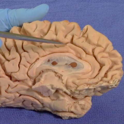
The Limbic System – Brain Dissections. The video demonstrates the Limbic System using 3D animations and gross specimens. Papez circuit is shown on gross specimens with mention of its involvement in memory. The role of the amygdala in fear and the olfactory cortex in temporal lobe epilepsy are described. The hippocampus from a brain with dementia is compared with a normal brain. With English or Italian closed captions. Video by Suzanne S. Stensaas, PhD, Professor Emeritus, Department of Neurobiology and Anatomy, University of Utah School of Medicine. Video retrieved from https://neurologicexam.med.utah.edu/adult/html/brain-dissections.html#26
Anatomical structures in item:
Uploaded by: M_Orsatti
Netherlands, Leiden – Leiden University Medical Center, Leiden University
Gyrus cinguli
Gyrus parahippocampalis
Corpus callosum
Truncus encephali
Hippocampus
Thalamus
Fornix
Hypothalamus
Pedunculus cerebri
Lobus temporalis
Commissura anterior
Uncus
Foramen interventriculare
Corpus mammillare
Corpus amygdaloideum
Encephalon
Creator(s)/credit: Professor Emeritus Suzanne S. Stensaas PhD
Requirements for usage
You are free to use this item if you follow the requirements of the license:  View license
View license If you use this item you should credit it as follows:
- For usage in print - copy and paste the line below:
- For digital usage (e.g. in PowerPoint, Impress, Word, Writer) - copy and paste the line below (optionally add the license icon):
"Utah NALab 26 - Brain Dissection Video The Limbic System - English and Italian closed captions" at AnatomyTOOL.org by Suzanne S. Stensaas, license: Creative Commons Attribution-NonCommercial-ShareAlike


Comments