
nid: 62665
Additional formats:
None available
Description:
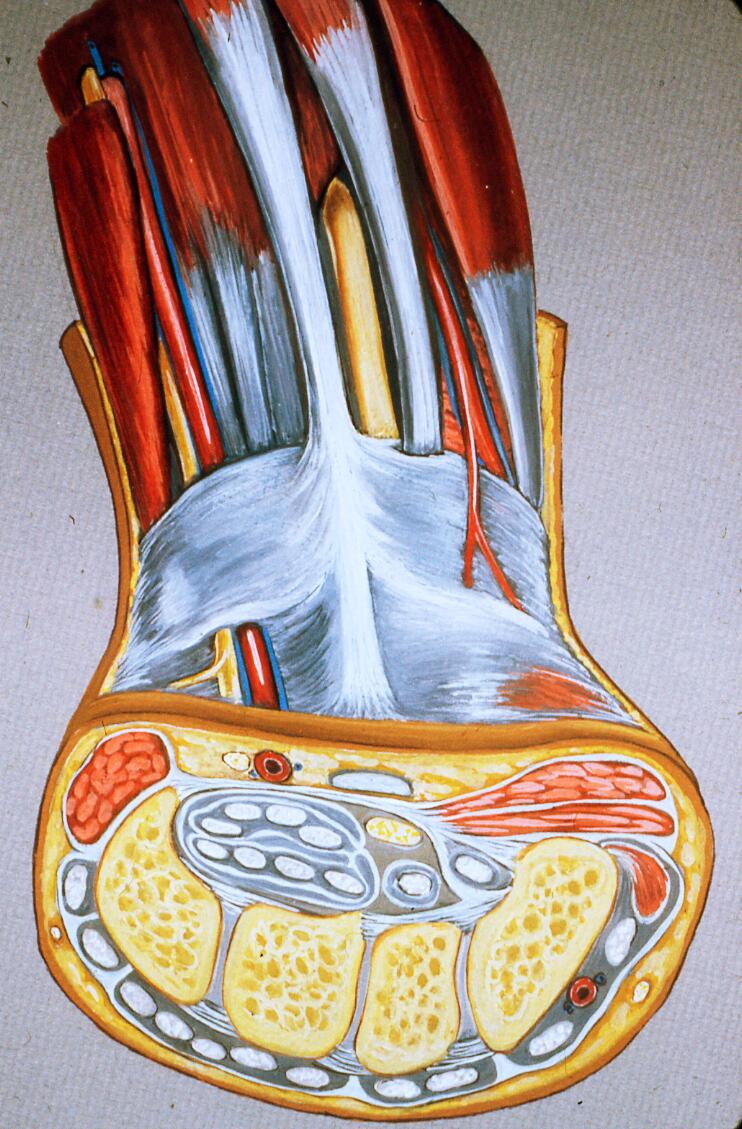
Muscles and tendons of hand. Artist's rendering* of dissected distal left forearm and wrist plus cross section of hand at level of the distal row of carpal bones, and flexor and extensor tendons.
*the cross sectional portion of the diagram is more diagramatic than the muscle/tendon portion.
Source: Orthopaedic Surgical Anatomy Teaching Collection (collection), Rehman and Smith Color Atlas of Orthopaedic Surgical Anatomy (subcollection), University of Southern California (contributing entity). Asset UC1570344.
Anatomical structures in item:
Uploaded by: rva
Netherlands, Leiden – Leiden University Medical Center, Leiden University
Canalis carpi
Antebrachium
Carpus
Manus
Os hamatum
Os trapezoideum
Os trapezium
Os capitatum
Nervus medianus
Retinaculum musculorum flexorum manus
Nervus ulnaris
Arteria radialis
Nervus ulnaris
Creator(s)/credit: Irving Rehman PhD, FICS; Chadwick F. Smith MD, PhD, FACS, FICS; Helen Barker
Requirements for usage
You are free to use this item if you follow the requirements of the license:  View license
View license If you use this item you should credit it as follows:
- For usage in print - copy and paste the line below:
- For digital usage (e.g. in PowerPoint, Impress, Word, Writer) - copy and paste the line below (optionally add the license icon):
"USC - Drawing Muscles and tendons of hand - no labels" at AnatomyTOOL.org by Irving Rehman, Chadwick F. Smith and Helen Barker, license: Creative Commons Attribution-NonCommercial-ShareAlike
"USC - Drawing Muscles and tendons of hand - no labels" by Irving Rehman, Chadwick F. Smith and Helen Barker, license: CC BY-NC-SA


Comments