
nid: 62671
Additional formats:
None available
Description:
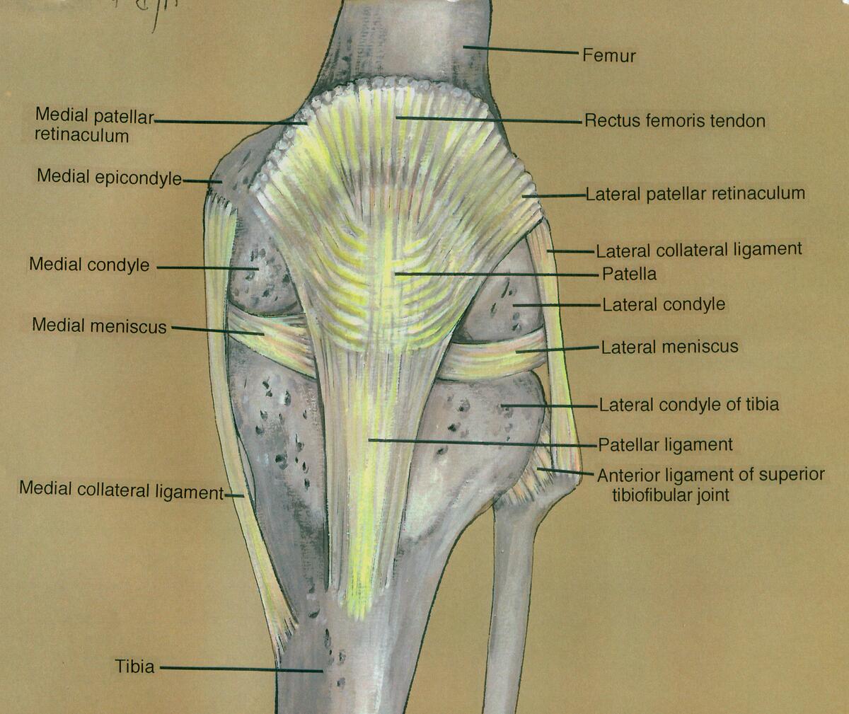
Knee, anterior view, showing patella, ligaments, menisci and bones. This drawing of the left knee is part of a series with different drawings on the anatomy of the knee.
Source: Norris Medical Library (collection), University of Southern California (contributing entity). Asset UC1569020.
Source: Norris Medical Library (collection), University of Southern California (contributing entity). Asset UC1569020.
Anatomical structures in item:
Uploaded by: rva
Netherlands, Leiden – Leiden University Medical Center, Leiden University
Genu
Femur
Tibia
Fibula
Condylus medialis femoris
Retinaculum patellae mediale
Epicondylus medialis femoris
Meniscus medialis
Ligamentum collaterale tibiale
Ligamentum capitis fibulae anterius
Superior tibiofibular joint
Ligamentum patellae
Patella
Condylus lateralis tibiae
Meniscus lateralis
Discus articularis
Condylus lateralis femoris
Ligamentum collaterale laterale articulationis talocruralis
Retinaculum patellae laterale
Creator(s)/credit: Irving Rehman PhD, FICS.; Chadwick F. Smith MD, PhD, FACS, FICS; Helen Barker
Requirements for usage
You are free to use this item if you follow the requirements of the license:  View license
View license If you use this item you should credit it as follows:
- For usage in print - copy and paste the line below:
- For digital usage (e.g. in PowerPoint, Impress, Word, Writer) - copy and paste the line below (optionally add the license icon):
"USC - Drawing Knee, anterior view, showing patella, ligaments, menisci and bones - English labels" at AnatomyTOOL.org by Irving Rehman, Chadwick F. Smith and Helen Barker, license: Creative Commons Attribution-NonCommercial-ShareAlike
"USC - Drawing Knee, anterior view, showing patella, ligaments, menisci and bones - English labels" by Irving Rehman, Chadwick F. Smith and Helen Barker, license: CC BY-NC-SA


Comments