nid: 63686
Additional formats:
None available
Description:
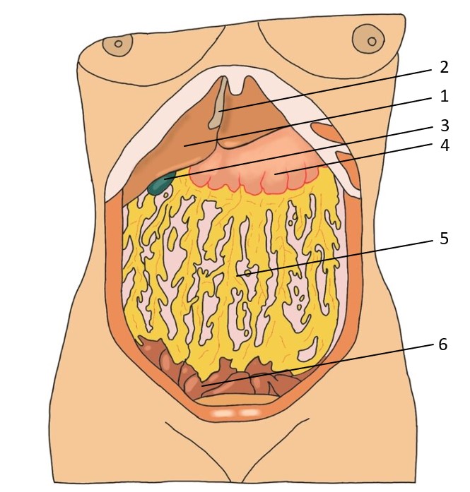
In this image we get an anterior view of the organs inside the peritoneal cavity after removal of the anterior abdominal wall and incision of the parietal peritoneum.
Labels: Hepar (1), Lig. falciforme hepatis (2), Vesica biliaris (3), Gaster (4), Omentum majus (5), Ileum (6)
Universiteit Gent - Students - Drawing Peritoneal cavity (2) - numbered Latin labels © 2023 by Manon Sierens is licensed under CC BY-NC-SA 4.0
Labels: Hepar (1), Lig. falciforme hepatis (2), Vesica biliaris (3), Gaster (4), Omentum majus (5), Ileum (6)
Universiteit Gent - Students - Drawing Peritoneal cavity (2) - numbered Latin labels © 2023 by Manon Sierens is licensed under CC BY-NC-SA 4.0
Anatomical structures in item:
Uploaded by: sehellin
Belgium, Gent (Ghent) - Ghent University
Hepar
Ligamentum falciforme hepatis
Vesica biliaris (Fellea)
Omentum majus
Ileum
Creator(s)/credit: Manon Sierens, student, Belgium, Gent (Ghent) - Ghent University; Jana Foulon, student, Belgium, Gent (Ghent) - Ghent University; Senne Hellinck, student, Belgium, Gent (Ghent) - Ghent University
Requirements for usage
You are free to use this item if you follow the requirements of the license:  View license
 View license
 If you use this item you should credit it as follows:
- For usage in print - copy and paste the line below:
- For digital usage (e.g. in PowerPoint, Impress, Word, Writer) - copy and paste the line below (optionally add the license icon):
"Universiteit Gent - Students - Drawing Peritoneal cavity (2) - numbered Latin labels" at AnatomyTOOL.org by Manon Sierens, Belgium, Gent (Ghent) - Ghent University, Jana Foulon, Belgium, Gent (Ghent) - Ghent University and Senne Hellinck, Belgium, Gent (Ghent) - Ghent University, license: Creative Commons Attribution-NonCommercial-ShareAlike
"Universiteit Gent - Students - Drawing Peritoneal cavity (2) - numbered Latin labels" by Manon Sierens, Belgium, Gent (Ghent) - Ghent University, Jana Foulon, Belgium, Gent (Ghent) - Ghent University and Senne Hellinck, Belgium, Gent (Ghent) - Ghent University, license: CC BY-NC-SA


Comments