
nid: 58902
Additional formats:
None available
Description:
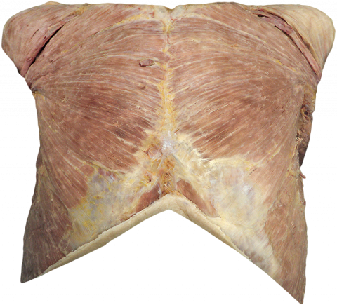
Anatomy of the thoracic wall. This image shows the thoracic wall with the skin removed. With the buttons on the right you can light up different anatomic structures.
Anatomical structures in item:
Uploaded by: rva
Netherlands, Leiden – Leiden University Medical Center, Leiden University
Thorax
Vena cephalica
Musculus deltoideus
Musculus obliquus externus abdominis
Arteria thoracica interna
Musculus pectoralis major
Creator(s)/credit: Dr Claudia Krebs, UBC; Monika Fejtek, UBC; Alexa Mordhorst, UBC
Requirements for usage
You are free to use this item if you follow the requirements of the license:  View license
View license If you use this item you should credit it as follows:
- For usage in print - copy and paste the line below:
- For digital usage (e.g. in PowerPoint, Impress, Word, Writer) - copy and paste the line below (optionally add the license icon):
"U.Br.Columbia - Photo Anatomy of the thoracic wall (dissection)" at AnatomyTOOL.org by Claudia Krebs, UBC, Monika Fejtek, UBC and Alexa Mordhorst, UBC, license: Creative Commons Attribution-NonCommercial-ShareAlike. Source: website Clinical Anatomy, http://www.clinicalanatomy.ca
"U.Br.Columbia - Photo Anatomy of the thoracic wall (dissection)" by Claudia Krebs, UBC, Monika Fejtek, UBC and Alexa Mordhorst, UBC, license: CC BY-NC-SA. Source: website Clinical Anatomy, http://www.clinicalanatomy.ca


Comments