
nid: 58911
Additional formats:
None available
Description:
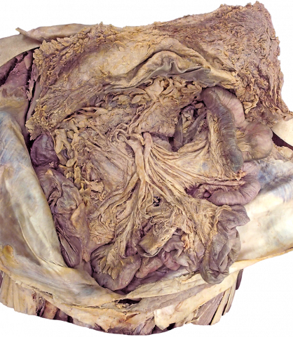
Anatomy of the midgut. In this image, the organs that belong to the midgut are visible. With the buttons on the right you can light up different anatomic structures.
Anatomical structures in item:
Uploaded by: rva
Netherlands, Leiden – Leiden University Medical Center, Leiden University
Colon ascendens
Appendices adiposae coli
Arteriae ileales
Vena ileocolica
Venae ileales
Arteria ileocolica
Ileum
Jejunum
Arteriae jejunales
Venae jejunales
Arteria marginalis coli
Arteria colica media
Vena colica media
Arteria colica dextra
Vena colica dextra
Vena mesenterica superior
Arteria mesenterica superior
Creator(s)/credit: Dr Claudia Krebs, UBC; Monika Fejtek, UBC; Alexa Mordhorst, UBC
Requirements for usage
You are free to use this item if you follow the requirements of the license:  View license
View license If you use this item you should credit it as follows:
- For usage in print - copy and paste the line below:
- For digital usage (e.g. in PowerPoint, Impress, Word, Writer) - copy and paste the line below (optionally add the license icon):
"U.Br.Columbia - Photo Anatomy of the midgut (dissection)" at AnatomyTOOL.org by Claudia Krebs, UBC, Monika Fejtek, UBC and Alexa Mordhorst, UBC, license: Creative Commons Attribution-NonCommercial-ShareAlike. Source: website Clinical Anatomy, http://www.clinicalanatomy.ca
"U.Br.Columbia - Photo Anatomy of the midgut (dissection)" by Claudia Krebs, UBC, Monika Fejtek, UBC and Alexa Mordhorst, UBC, license: CC BY-NC-SA. Source: website Clinical Anatomy, http://www.clinicalanatomy.ca


Comments