
nid: 59568
Additional formats:
None available
Description:
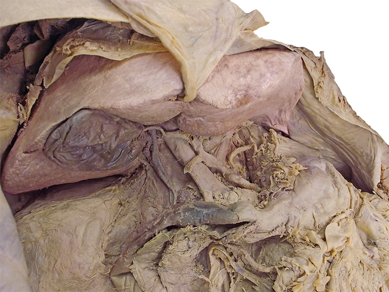
Anatomy of the foregut. In this image, the organs that belong to the foregut are visible. No labels.
Retrieved from website Clinical Anatomy of the University of British Columbia.
Retrieved from website Clinical Anatomy of the University of British Columbia.
Anatomical structures in item:
Uploaded by: rva
Netherlands, Leiden – Leiden University Medical Center, Leiden University
Abdomen
Duodenum
Ligamentum falciforme hepatis
Vesica biliaris (Fellea)
Hepar
Pancreas
Ventriculus
Creator(s)/credit: Prof. Claudia Krebs MD, PhD, anatomist, UBC; Monika Fejtek, digital media technologist, UBC; Alexa Mordhorst MD, UBC
Requirements for usage
You are free to use this item if you follow the requirements of the license:  View license
View license If you use this item you should credit it as follows:
- For usage in print - copy and paste the line below:
- For digital usage (e.g. in PowerPoint, Impress, Word, Writer) - copy and paste the line below (optionally add the license icon):
"U.Br.Columbia - Photo Anatomy of the foregut - no labels (dissection)" at AnatomyTOOL.org by Claudia Krebs, UBC, Monika Fejtek, UBC and Alexa Mordhorst, UBC, license: Creative Commons Attribution-NonCommercial-ShareAlike. Source: website Clinical Anatomy, http://www.clinicalanatomy.ca
"U.Br.Columbia - Photo Anatomy of the foregut - no labels (dissection)" by Claudia Krebs, UBC, Monika Fejtek, UBC and Alexa Mordhorst, UBC, license: CC BY-NC-SA. Source: website Clinical Anatomy, http://www.clinicalanatomy.ca


Comments