
nid: 60423
Additional formats:
None available
Description:
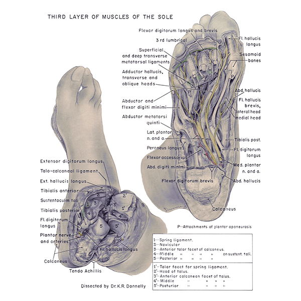
Third layer of muscles of the sole. The third layer of muscles of the sole are visible in the right image. In the left image, the anatomy of the calcaneus and talus is visualized. English labels.
Retrieved from website Clinical Anatomy of the University of British Columbia.
Retrieved from website Clinical Anatomy of the University of British Columbia.
Anatomical structures in item:
Uploaded by: rva
Netherlands, Leiden – Leiden University Medical Center, Leiden University
Pes
Ligamentum calcaneonaviculare plantare
Os scaphoideum
Calcaneus
Facies articularis talaris media calcanei
Facies articularis talaris anterior calcanei
Facies articularis talaris posterior calcanei
Caput tali
Facies articularis calcanea anterior tali
Facies articularis calcanea media tali
Facies articularis calcanea posterior tali
Musculus lumbricales pedis
Os sesamoideum
Requirements for usage
You are free to use this item if you follow the requirements of the license:  View license
View license If you use this item you should credit it as follows:
- For usage in print - copy and paste the line below:
- For digital usage (e.g. in PowerPoint, Impress, Word, Writer) - copy and paste the line below (optionally add the license icon):
"U.Br.Columbia - Drawing Third layer of muscles of the sole - English labels" at AnatomyTOOL.org by , license: Creative Commons Attribution-NonCommercial-ShareAlike. Created for: Department of Anatomy (now Department of Cellular and Physiological Sciences) at the University of British Columbia. Source: website Clinical Anatomy, http://www.clinicalanatomy.ca
"U.Br.Columbia - Drawing Third layer of muscles of the sole - English labels" by , license: CC BY-NC-SA. Created for: Department of Anatomy (now Department of Cellular and Physiological Sciences) at the University of British Columbia. Source: website Clinical Anatomy, http://www.clinicalanatomy.ca


Comments