
nid: 59899
Additional formats:
None available
Description:
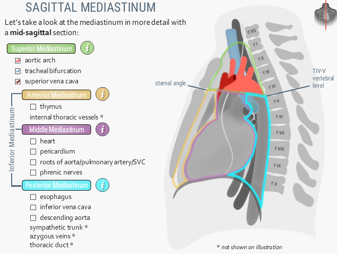
The superior mediastinum. This image shows a sagittal view of the mediastinum. The superior, anterior, middle and posterior mediastinum are marked with respectively a green, orange, purple and light blue line. The superior mediastinum contains the aortic arch, tracheal bifurcation and the superior vena cava. English labels. Retrieved from the interactive module Around the heart: Mediastinum from the website Clinical Anatomy of the University of British Columbia.
Anatomical structures in item:
Uploaded by: rva
Netherlands, Leiden – Leiden University Medical Center, Leiden University
Cor
Mediastinum
Mediastinum superius
Cavitas thoracis
Arcus aortae
Vena cava superior
Bifurcatio tracheae
Creator(s)/credit: Prof. Claudia Krebs MD, PhD, anatomist, UBC; Monika Fejtek, digital media technologist, UBC; Rebecca Comeau MD, UBC; Dr. Olusegun Oyedele; Dr. Paul Rea; Daniel McClusky; Iskander Afiq Mohamad Hashim; Jenna Woods
Requirements for usage
You are free to use this item if you follow the requirements of the license:  View license
View license If you use this item you should credit it as follows:
- For usage in print - copy and paste the line below:
- For digital usage (e.g. in PowerPoint, Impress, Word, Writer) - copy and paste the line below (optionally add the license icon):
"U.Br.Columbia - Drawing The superior mediastinum - English labels" at AnatomyTOOL.org by Claudia Krebs, UBC, Monika Fejtek, UBC, Rebecca Comeau, UBC et al, license: Creative Commons Attribution-NonCommercial-ShareAlike. Source: website Clinical Anatomy, http://www.clinicalanatomy.ca
"U.Br.Columbia - Drawing The superior mediastinum - English labels" by Claudia Krebs, UBC, Monika Fejtek, UBC, Rebecca Comeau, UBC et al, license: CC BY-NC-SA. Source: website Clinical Anatomy, http://www.clinicalanatomy.ca


Comments