
nid: 60395
Additional formats:
None available
Description:
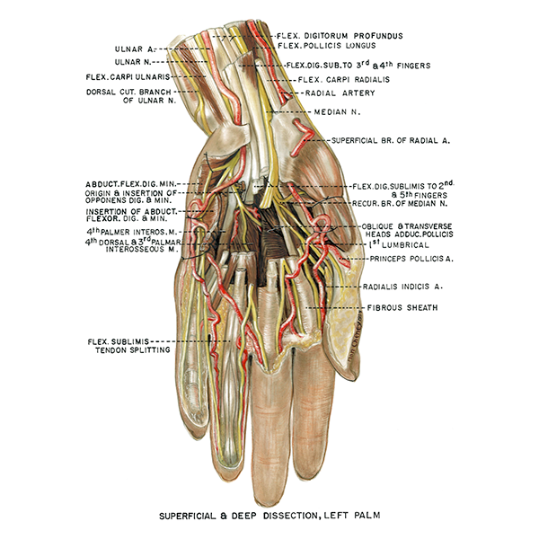
Superficial and deep dissection of left palm. Both the superficial and deep muscles can be seen in this image. English labels.
Retrieved from website Clinical Anatomy of the University of British Columbia.
Retrieved from website Clinical Anatomy of the University of British Columbia.
Anatomical structures in item:
Uploaded by: rva
Netherlands, Leiden – Leiden University Medical Center, Leiden University
Manus
Arteria ulnaris
Nervus ulnaris
Musculus flexor carpi ulnaris
Musculi interossei palmares
Vaginae fibrosae digitorum manus
Arteria radialis indicis
Arteria princeps pollicis
Musculi lumbricales manus
Nervus medianus
Creator(s)/credit: A.G.L. (Nan) Cheney, medical illustrator, UBC
Requirements for usage
You are free to use this item if you follow the requirements of the license:  View license
View license If you use this item you should credit it as follows:
- For usage in print - copy and paste the line below:
- For digital usage (e.g. in PowerPoint, Impress, Word, Writer) - copy and paste the line below (optionally add the license icon):
"U.Br.Columbia - Drawing Superficial and deep dissection of left palm - English labels" at AnatomyTOOL.org by A.G.L. (Nan) Cheney, UBC, license: Creative Commons Attribution-NonCommercial-ShareAlike. Source: website Clinical Anatomy, http://www.clinicalanatomy.ca
"U.Br.Columbia - Drawing Superficial and deep dissection of left palm - English labels" by A.G.L. (Nan) Cheney, UBC, license: CC BY-NC-SA. Source: website Clinical Anatomy, http://www.clinicalanatomy.ca


Comments