nid: 60417
Additional formats:
None available
Description:
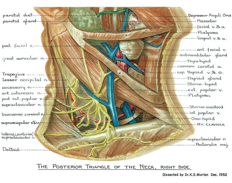
The posterior triangle of the neck. The structures of the posterior triangle of the neck are shown. Also, a part of the anterior triangle are shown. English labels.
Retrieved from website Clinical Anatomy of the University of British Columbia.
Retrieved from website Clinical Anatomy of the University of British Columbia.
Anatomical structures in item:
Uploaded by: rva
Netherlands, Leiden – Leiden University Medical Center, Leiden University
Ductus parotideus
Trigonum cervicale posterius
Musculus trapezius
Glandula parotidea
Vena facialis
Nervus auricularis magnus
Nervus occipitalis minor
Nervus accessorius [XI]
Nervus transversus cervicalis
Vena jugularis externa
Nervi supraclaviculares
Arteria transversa cervicis
Nervus suprascapularis
Arteria suprascapularis
Nervi supraclaviculares laterales
Nervi supraclaviculares
Clavicula
Musculus omohyoideus
Vena jugularis interna
Musculus sternocleidomastoideus
Platysma
Musculus sternohyoideus
Glandula thyroidea
Vena thyroidea superior
Arteria thyroidea superior
Arteria carotis communis
Musculus thyrohyoideus
Glandula submandibularis
Vena facialis
Vena retromandibularis
Vena lingualis
Arteria lingualis
Arteria facialis
Requirements for usage
You are free to use this item if you follow the requirements of the license:  View license
 View license
 If you use this item you should credit it as follows:
- For usage in print - copy and paste the line below:
- For digital usage (e.g. in PowerPoint, Impress, Word, Writer) - copy and paste the line below (optionally add the license icon):
"U.Br.Columbia - Drawing The posterior triangle of the neck - English labels" at AnatomyTOOL.org by , license: Creative Commons Attribution-NonCommercial-ShareAlike. Source: website Clinical Anatomy, http://www.clinicalanatomy.ca
"U.Br.Columbia - Drawing The posterior triangle of the neck - English labels" by , license: CC BY-NC-SA. Source: website Clinical Anatomy, http://www.clinicalanatomy.ca


Comments