
nid: 60359
Additional formats:
None available
Description:
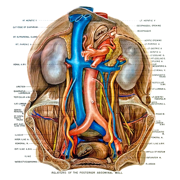
Nerves and vessels of the posterior abdominal wall. The relations of the veins, arteries and nerves of the posterior abdominal wall to the muscles and other structures is shown. English labels.
Retrieved from website Clinical Anatomy of the University of British Columbia.
Retrieved from website Clinical Anatomy of the University of British Columbia.
Anatomical structures in item:
Uploaded by: rva
Netherlands, Leiden – Leiden University Medical Center, Leiden University
Arteria iliaca communis
Vena hepatica dextra
Glandula suprarenalis
Arteria renalis
Ureter
Musculus quadratus lumborum
Arteria testicularis
Vena testicularis
Ligamentum iliolumbale
Nervus genitofemoralis
Spina iliaca anterior superior
Arteria iliaca interna
Nervus femoralis
Vena iliaca externa
Arteria iliaca externa
Vesica urinaria
Nervus obturatorius
Nervus iliohypogastricus
Musculus psoas major
Arteria mesenterica inferior
Nervus genitofemoralis
Vena renalis sinistra
Vena renalis dextra
Venae renales
Abdomen
Arteria mesenterica superior
Arteria lienalis
Arteria hepatica
Arteria gastrica sinistra
Arteria phrenica inferior
Aorta
Aorta abdominalis
Aorta descendens
Bifurcatio aortae
Constrictio diaphragmatica oesophageae
Vena hepatica sinistra
Creator(s)/credit: A.G.L. (Nan) Cheney, medical illustrator, UBC
Requirements for usage
You are free to use this item if you follow the requirements of the license:  View license
View license If you use this item you should credit it as follows:
- For usage in print - copy and paste the line below:
- For digital usage (e.g. in PowerPoint, Impress, Word, Writer) - copy and paste the line below (optionally add the license icon):
"U.Br.Columbia - Drawing Nerves and vessels of the posterior abdominal wall - English labels" at AnatomyTOOL.org by A.G.L. (Nan) Cheney, UBC, license: Creative Commons Attribution-NonCommercial-ShareAlike. Source: website Clinical Anatomy, http://www.clinicalanatomy.ca
"U.Br.Columbia - Drawing Nerves and vessels of the posterior abdominal wall - English labels" by A.G.L. (Nan) Cheney, UBC, license: CC BY-NC-SA. Source: website Clinical Anatomy, http://www.clinicalanatomy.ca


Comments