
nid: 59873
Additional formats:
None available
Description:
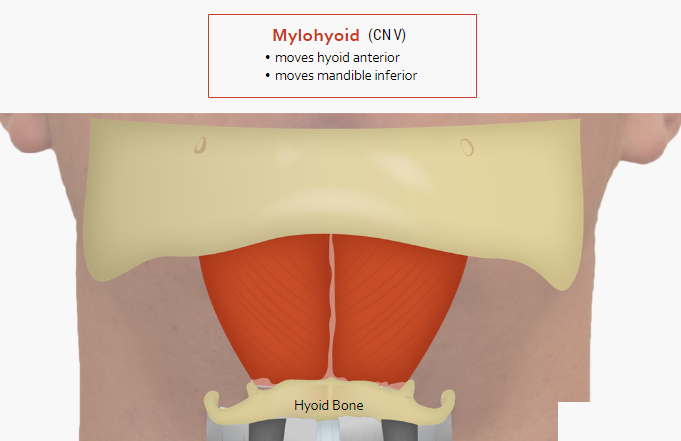
Mylohyoid muscle. In this image, the anatomy of the mylohyoid muscle can be appreciated. This suprahyoid muscle lies over the geniohyoid muscle and is innervated by the trigeminal nerve (CN V). English labels. Retrieved from the interactive module Anatomy of swallowing (Deglutition) from the website Clinical Anatomy of the University of British Columbia.
Anatomical structures in item:
Uploaded by: rva
Netherlands, Leiden – Leiden University Medical Center, Leiden University
Musculus mylohyoideus
Os hyoideum
Musculi suprahyoidei
Mandibula
Creator(s)/credit: Prof. Claudia Krebs MD, PhD, anatomist, UBC; Monika Fejtek, digital media technologist, UBC; Stacey Skoretz, UBC; Stephanie Riopelle, UBC; Veronica Letawski, UBC; Ajay Grewal, UBC; Paige Blumer, UBC; Connor Dunne, UBC; Curtis J. Logan, UBC; Mark Dykstra, UBC
Requirements for usage
You are free to use this item if you follow the requirements of the license:  View license
View license If you use this item you should credit it as follows:
- For usage in print - copy and paste the line below:
- For digital usage (e.g. in PowerPoint, Impress, Word, Writer) - copy and paste the line below (optionally add the license icon):
"U.Br.Columbia - Drawing Mylohyoid muscle - English labels" at AnatomyTOOL.org by Claudia Krebs, UBC, Monika Fejtek, UBC, Stacey Skoretz, UBC et al, license: Creative Commons Attribution-NonCommercial-ShareAlike. Source: website Clinical Anatomy, http://www.clinicalanatomy.ca
"U.Br.Columbia - Drawing Mylohyoid muscle - English labels" by Claudia Krebs, UBC, Monika Fejtek, UBC, Stacey Skoretz, UBC et al, license: CC BY-NC-SA. Source: website Clinical Anatomy, http://www.clinicalanatomy.ca


Comments