
nid: 60142
Additional formats:
None available
Description:
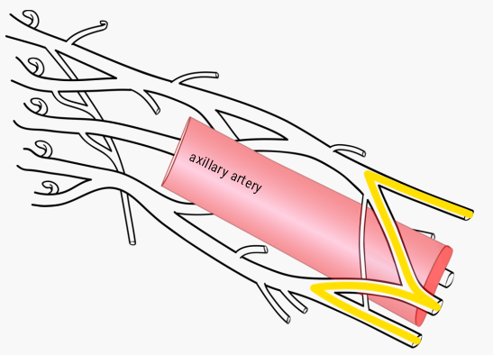
The "M" landmark of the brachial plexus. Upon identifying the "M" shape (here depicted in yellow), the cords, branches and terminal nerves of the brachial plexus can be identified easily. The "M" is formed by the branches as they recombine into the terminal nerves, this "M" wraps neatly around the axillary artery. No labels. Retrieved from the interactive module Brachial plexus from the website Clinical Anatomy of the University of British Columbia.
Anatomical structures in item:
Uploaded by: rva
Netherlands, Leiden – Leiden University Medical Center, Leiden University
Plexus brachialis
Arteria axillaris
Creator(s)/credit: Prof. Claudia Krebs MD, PhD, anatomist, UBC; Monika Fejtek, digital media technologist, UBC; Megan Leong, medical student, UBC; Mackenzie Parry, medical student; Paige Blumer, medical illustrator; Connor Dunne; Dr Timothy Inglis
Requirements for usage
You are free to use this item if you follow the requirements of the license:  View license
View license If you use this item you should credit it as follows:
- For usage in print - copy and paste the line below:
- For digital usage (e.g. in PowerPoint, Impress, Word, Writer) - copy and paste the line below (optionally add the license icon):
"U.Br.Columbia - Drawing The "M" landmark of the brachial plexus - English labels" at AnatomyTOOL.org by Claudia Krebs, UBC, Monika Fejtek, UBC, Megan Leong, UBC et al, license: Creative Commons Attribution-NonCommercial-ShareAlike. Source: website Clinical Anatomy, http://www.clinicalanatomy.ca
"U.Br.Columbia - Drawing The "M" landmark of the brachial plexus - English labels" by Claudia Krebs, UBC, Monika Fejtek, UBC, Megan Leong, UBC et al, license: CC BY-NC-SA. Source: website Clinical Anatomy, http://www.clinicalanatomy.ca


Comments