
nid: 59905
Additional formats:
None available
Description:
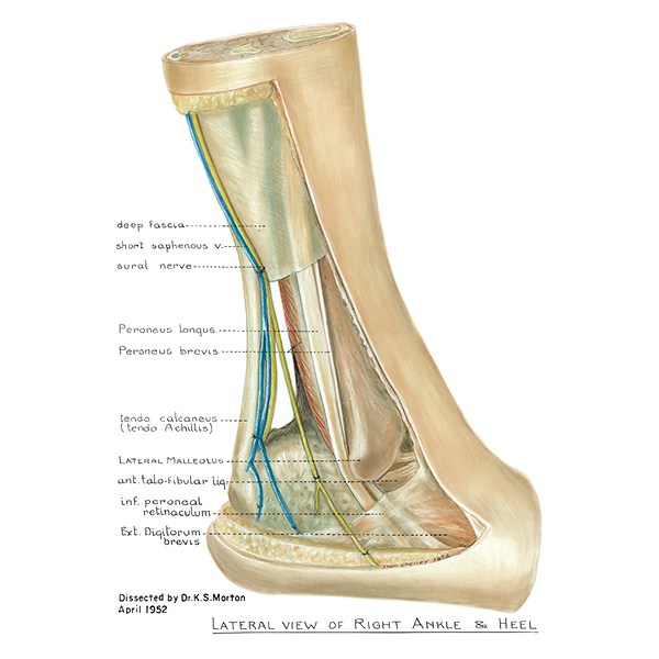
Lateral view of right ankle and heel. The lateral ankle and heel is shown in this image. English labels.
Retrieved from website Clinical Anatomy of the University of British Columbia.
Retrieved from website Clinical Anatomy of the University of British Columbia.
Anatomical structures in item:
Uploaded by: rva
Netherlands, Leiden – Leiden University Medical Center, Leiden University
Tarsus
Calx
Fascia cruris
Vena saphena parva
Nervus suralis
Musculus fibularis longus
Musculus fibularis brevis
Tendo calcaneus
Malleolus lateralis
Ligamentum talofibulare anterius
Retinaculum musculorum fibularium inferius
Musculus extensor digitorum brevis
Creator(s)/credit: A.G.L. (Nan) Cheney, medical illustrator, UBC; Dr. K.S. Morton, UBC
Requirements for usage
You are free to use this item if you follow the requirements of the license:  View license
View license If you use this item you should credit it as follows:
- For usage in print - copy and paste the line below:
- For digital usage (e.g. in PowerPoint, Impress, Word, Writer) - copy and paste the line below (optionally add the license icon):
"U.Br.Columbia - Drawing Lateral view of right ankle and heel - English labels" at AnatomyTOOL.org by A.G.L. (Nan) Cheney, UBC and K.S. Morton, UBC, license: Creative Commons Attribution-NonCommercial-ShareAlike. Source: website Clinical Anatomy, http://www.clinicalanatomy.ca
"U.Br.Columbia - Drawing Lateral view of right ankle and heel - English labels" by A.G.L. (Nan) Cheney, UBC and K.S. Morton, UBC, license: CC BY-NC-SA. Source: website Clinical Anatomy, http://www.clinicalanatomy.ca


Comments