nid: 62521
Additional formats:
None available
Description:
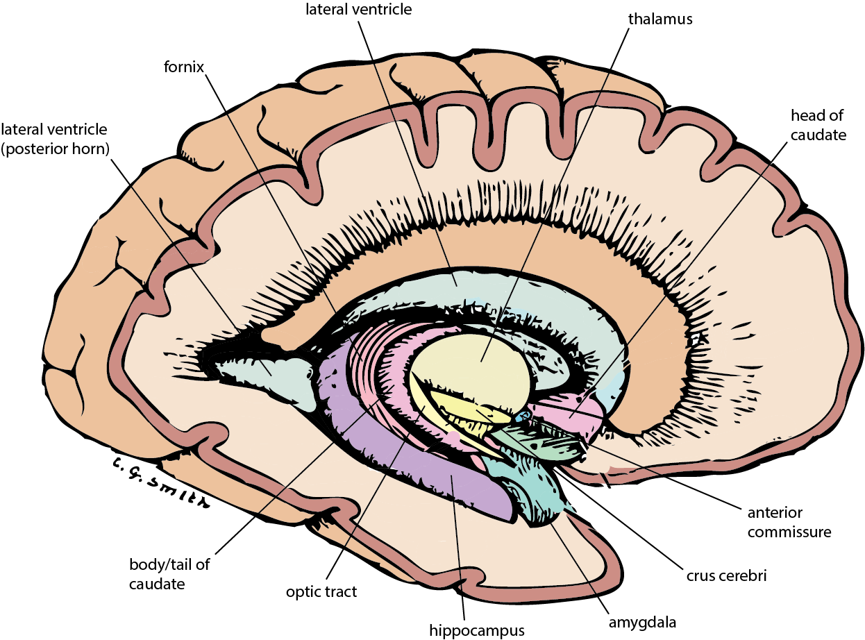
Lateral view of brain: basal ganglia (medial). This image shows the most medial structures of the basal ganglia. English labels.
Retrieved from www.neuroanatomy.ca.
Retrieved from www.neuroanatomy.ca.
Anatomical structures in item:
Uploaded by: rva
Netherlands, Leiden – Leiden University Medical Center, Leiden University
Fornix
Pars centralis ventriculi lateralis
Thalamus
Caput nuclei caudati
Nucleus caudatus
Commissura anterior
Crus cerebri
Hippocampus
Corpus amygdaloideum
Tractus opticus
Corpus nuclei caudati
Cauda nuclei caudati
Creator(s)/credit: C.G. Smith
Requirements for usage
You are free to use this item if you follow the requirements of the license:  View license
 View license
 If you use this item you should credit it as follows:
- For usage in print - copy and paste the line below:
- For digital usage (e.g. in PowerPoint, Impress, Word, Writer) - copy and paste the line below (optionally add the license icon):
"U.Br.Columbia - Drawing Lateral view of brain: basal ganglia (medial) - English labels" at AnatomyTOOL.org by C.G. Smith, license: Creative Commons Attribution-NonCommercial-ShareAlike
"U.Br.Columbia - Drawing Lateral view of brain: basal ganglia (medial) - English labels" by C.G. Smith, license: CC BY-NC-SA


Comments