
nid: 62520
Additional formats:
None available
Description:
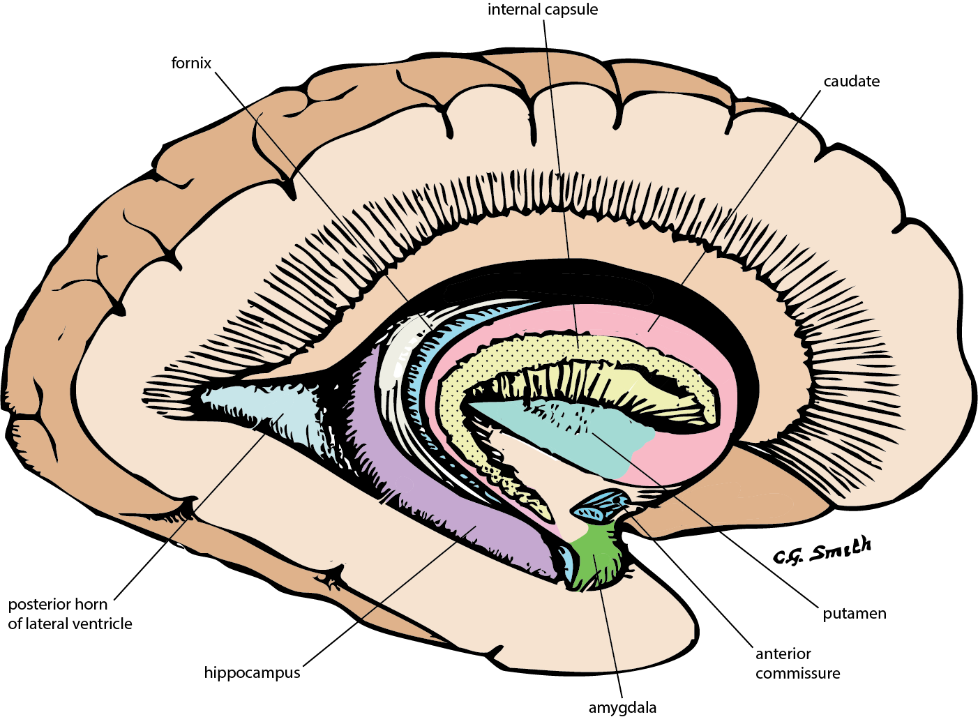
Lateral view of brain: basal ganglia (lateral). This image shows the most lateral structures of the basal ganglia. English labels.
Retrieved from www.neuroanatomy.ca.
Retrieved from www.neuroanatomy.ca.
Anatomical structures in item:
Uploaded by: rva
Netherlands, Leiden – Leiden University Medical Center, Leiden University
Fornix
Encephalon
Capsula interna
Nucleus caudatus
Putamen
Commissura anterior
Corpus amygdaloideum
Hippocampus
Cornu occipitale ventriculi lateralis
Creator(s)/credit: C.G. Smith
Requirements for usage
You are free to use this item if you follow the requirements of the license:  View license
View license If you use this item you should credit it as follows:
- For usage in print - copy and paste the line below:
- For digital usage (e.g. in PowerPoint, Impress, Word, Writer) - copy and paste the line below (optionally add the license icon):
"U.Br.Columbia - Drawing Lateral view of brain: basal ganglia (lateral) - English labels" at AnatomyTOOL.org by C.G. Smith, license: Creative Commons Attribution-NonCommercial-ShareAlike
"U.Br.Columbia - Drawing Lateral view of brain: basal ganglia (lateral) - English labels" by C.G. Smith, license: CC BY-NC-SA


Comments