
nid: 59743
Additional formats:
None available
Description:
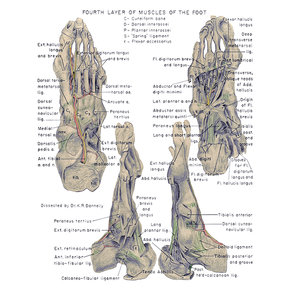
Fourth layer of muscles of the sole. The fourth layer of muscles of the sole is visible in this figure, the muscles can be appreciated in multiple directions. English labels.
Retrieved from website Clinical Anatomy of the University of British Columbia.
Retrieved from website Clinical Anatomy of the University of British Columbia.
Anatomical structures in item:
Uploaded by: rva
Netherlands, Leiden – Leiden University Medical Center, Leiden University
Pes
Planta
Musculus extensor hallucis longus
Musculus extensor hallucis brevis
Ligamenta tarsi dorsalia
Ligamentum cuneonavicularia dorsalia
Arteria dorsalis pedis
Arteria tibialis anterior
Os cuneiforme mediale
Os cuneiforme intermedium
Os cuneiforme laterale
Rami malleolares laterales (Arteria fibularis)
Arteria arcuata
Musculus fibularis tertius
Ligamentum calcaneofibulare
Ligamentum talocalcaneum posterius
Ligamentum collaterale mediale articulationis talocruralis
Creator(s)/credit: Department of Anatomy, University of British Columbia, UBC; Dr. K.R. Donnelly, UBC
Requirements for usage
You are free to use this item if you follow the requirements of the license:  View license
View license If you use this item you should credit it as follows:
- For usage in print - copy and paste the line below:
- For digital usage (e.g. in PowerPoint, Impress, Word, Writer) - copy and paste the line below (optionally add the license icon):
"U.Br.Columbia - Drawing Fourth layer of muscles of the sole - English labels" at AnatomyTOOL.org by Department of Anatomy, University of British Columbia, UBC and K.R. Donnelly, UBC, license: Creative Commons Attribution-NonCommercial-ShareAlike. Created for: Department of Anatomy (now Department of Cellular and Physiological Sciences) at the University of British Columbia. Source: website Clinical Anatomy, http://www.clinicalanatomy.ca
"U.Br.Columbia - Drawing Fourth layer of muscles of the sole - English labels" by Department of Anatomy, University of British Columbia, UBC and K.R. Donnelly, UBC, license: CC BY-NC-SA. Created for: Department of Anatomy (now Department of Cellular and Physiological Sciences) at the University of British Columbia. Source: website Clinical Anatomy, http://www.clinicalanatomy.ca


Comments