
nid: 59688
Additional formats:
None available
Description:
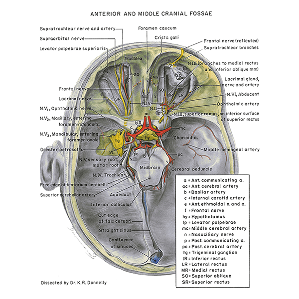
Anterior and middle cranial fossae. This figure shows the anterior and middle cranial fossae and their penetrating structures. English labels.
Retrieved from website Clinical Anatomy of the University of British Columbia.
Retrieved from website Clinical Anatomy of the University of British Columbia.
Anatomical structures in item:
Uploaded by: rva
Netherlands, Leiden – Leiden University Medical Center, Leiden University
Caput
Foramen caecum
Arteria meningea media
Pedunculus cerebri
Sinus rectus
Arteria cerebri anterior
Arteria cerebri posterior
Foramen rotundum
Ganglion trigeminale
Foramen ovale
Nervus opticus
Nervus oculomotorius [III]
Basilaris
Nervus petrosus major
Creator(s)/credit: Department of Anatomy, University of British Columbia, UBC; Dr K.R. Donnelly, anatomist, UBC
Requirements for usage
You are free to use this item if you follow the requirements of the license:  View license
View license If you use this item you should credit it as follows:
- For usage in print - copy and paste the line below:
- For digital usage (e.g. in PowerPoint, Impress, Word, Writer) - copy and paste the line below (optionally add the license icon):
"U.Br.Columbia - Drawing Anterior and middle cranial fossae - English labels" at AnatomyTOOL.org by Department of Anatomy, University of British Columbia, UBC and K.R. Donnelly, UBC, © Created for: Department of Anatomy (now Department of Cellular and Physiological Sciences) at the University of British Columbia , license: Creative Commons Attribution-NonCommercial-ShareAlike. Source: website Clinical Anatomy, http://www.clinicalanatomy.ca
"U.Br.Columbia - Drawing Anterior and middle cranial fossae - English labels" by Department of Anatomy, University of British Columbia, UBC and K.R. Donnelly, UBC, © Created for: Department of Anatomy (now Department of Cellular and Physiological Sciences) at the University of British Columbia , license: CC BY-NC-SA. Source: website Clinical Anatomy, http://www.clinicalanatomy.ca


Comments