
nid: 61943
Additional formats:
None available
Description:
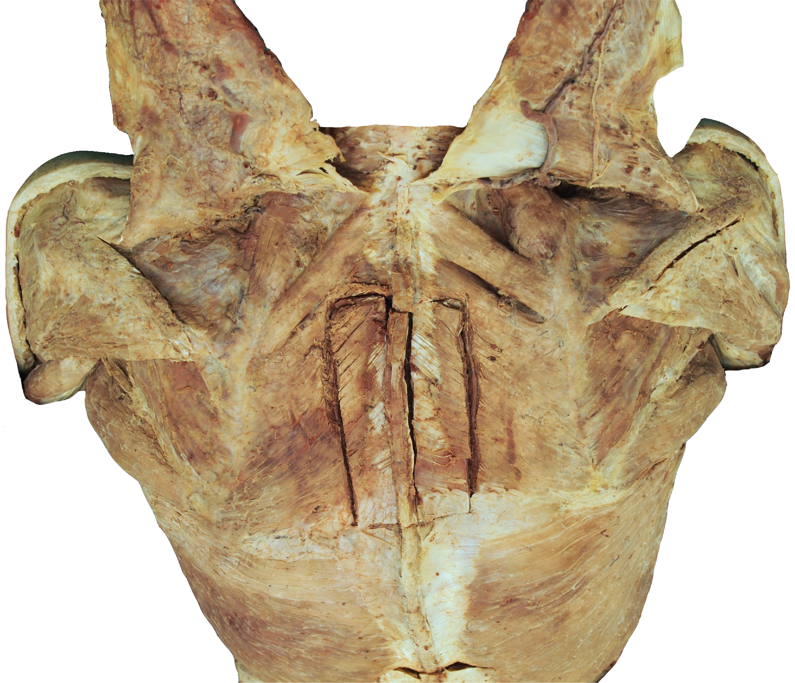
Deep back muscles. In this dissection image, the deep muscles of the back can be seen from posterior. The trapezius muscle is reflected, to make the accessory nerve visible. Structures can be highlighted on www.clinicalanatomy.ca.
Retrieved from website Clinical Anatomy of the University of British Columbia.
Retrieved from website Clinical Anatomy of the University of British Columbia.
Anatomical structures in item:
Uploaded by: rva
Netherlands, Leiden – Leiden University Medical Center, Leiden University
Dorsum
Nervus accessorius [XI]
Musculus deltoideus
Musculus infraspinatus
Musculus latissimus dorsi
Musculus levator scapulae
Musculus rhomboideus major
Musculus rhomboideus minor
Margo medialis scapulae
Acromion
Spina scapulae
Musculus supraspinatus
Musculus teres major
Musculus teres minor
Arteria transversa cervicis
Musculus trapezius
Creator(s)/credit: Prof. Claudia Krebs MD, PhD, anatomist, UBC; Monika Fejtek, digital media technologist, UBC; Alexa Mordhorst MD, UBC
Requirements for usage
You are free to use this item if you follow the requirements of the license:  View license
View license If you use this item you should credit it as follows:
- For usage in print - copy and paste the line below:
- For digital usage (e.g. in PowerPoint, Impress, Word, Writer) - copy and paste the line below (optionally add the license icon):
"U.Br.Columbia - Dissection Deep back muscles - no labels" at AnatomyTOOL.org by Claudia Krebs, UBC, Monika Fejtek, UBC and Alexa Mordhorst, UBC, license: Creative Commons Attribution-NonCommercial-ShareAlike. Source: website Clinical Anatomy, http://www.clinicalanatomy.ca
"U.Br.Columbia - Dissection Deep back muscles - no labels" by Claudia Krebs, UBC, Monika Fejtek, UBC and Alexa Mordhorst, UBC, license: CC BY-NC-SA. Source: website Clinical Anatomy, http://www.clinicalanatomy.ca


Comments