
nid: 59192
Additional formats:
None available
Description:
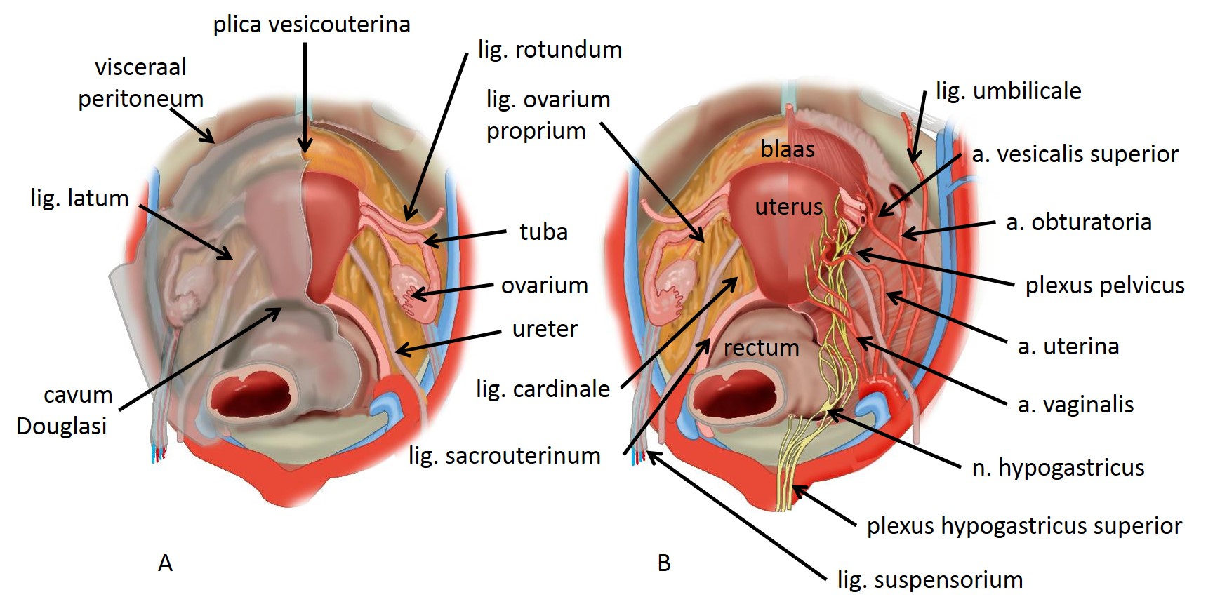
Superior view of female pelvis viscera, arterial supply and pelvic plexus. On the left (A) the peritoneum covering the pelvic viscera is seen, including the broad ligament of the uterus. The peritoneal pouches; cavum Douglasi and vesico-uterine pouch. On the right halve of the left: peritoneum has been removed; the round ligament of the uterus and the ovarian ligament are shown. On the right (B) the bladder, uterus and rectum are labeled. On the right halve the corpus intrapelvinum has been dissected, showing the pelvic plexus and arteries. Dutch labels.
Illustration by Ron Slagter and Marco DeRuiter for course 'Surgical Anatomy of the lesser pelvis' by the 'Urologisch Opleidings Instituut', the Netherlands.
Illustration by Ron Slagter and Marco DeRuiter for course 'Surgical Anatomy of the lesser pelvis' by the 'Urologisch Opleidings Instituut', the Netherlands.
Anatomical structures in item:
Uploaded by: Siem Zethof
Netherlands, Leiden – Leiden University Medical Center, Leiden University
Ligamentum latum uteri
Excavatio rectouterina
Ureter
Uterus
Ovarium
Tuba uterina (Salpinx)
Ligamentum teres uteri
Ligamentum ovarii proprium
Ligamentum cardinale
Ligamentum rectouterinum
Plexus nervosus hypogastricus inferior
Arteria uterina
Arteria obturatoria
Pelvis
Creator(s)/credit: Ron Slagter NZIMBI, medical illustrator, LUMC; Prof. Marco DeRuiter PhD, anatomist, LUMC
Requirements for usage
You are free to use this item if you follow the requirements of the license:  View license
View license If you use this item you should credit it as follows:
- For usage in print - copy and paste the line below:
- For digital usage (e.g. in PowerPoint, Impress, Word, Writer) - copy and paste the line below (optionally add the license icon):
"Superior view of female pelvis viscera, peritoneum, arterial supply and pelvic plexus – Dutch labels" at AnatomyTOOL.org by Ron Slagter, LUMC and Marco DeRuiter, LUMC, license: Creative Commons Attribution-NonCommercial-ShareAlike


Comments