
nid: 62321
Additional formats:
None available
Description:
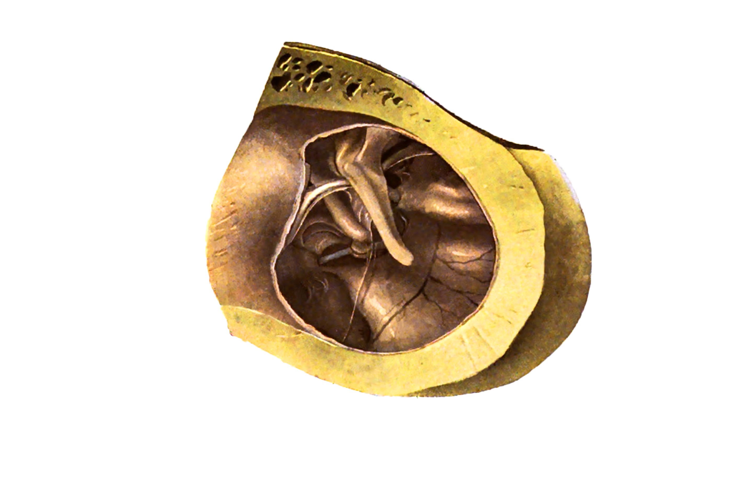
Outer wall of the right tympanic cavity, lateral view.
From 'Atlas and Textbook of Human Anatomy', 1911 (?), Vol. 3, fig.807, by Johannes Sobotta and J. Playfair McMurrich. Artist: K. Hajek. Retrieved from Sobotta's Anatomy plates at Wikimedia.
Image editing by dream_studio3.
From 'Atlas and Textbook of Human Anatomy', 1911 (?), Vol. 3, fig.807, by Johannes Sobotta and J. Playfair McMurrich. Artist: K. Hajek. Retrieved from Sobotta's Anatomy plates at Wikimedia.
Image editing by dream_studio3.
Anatomical structures in item:
Uploaded by: rva
Netherlands, Leiden – Leiden University Medical Center, Leiden University
Cavitas tympani
Spina tympanica major
Spina tympanica minor
Malleus
Chorda tympani
Plica mallearis anterior
Musculus tensor tympani
Musculus tensor tympani
Promontorium tympani
Stapes
Processus lenticularis incudis
Membrana tympanica
Chorda tympani
Incus
Meatus acusticus externus
Eminentia pyramidalis
Fossula fenestrae cochleae
Collum mallei
Crus longum incudis
Creator(s)/credit: Prof.dr. Johannes Sobotta, anatomist; dream_studio3 BA, image editing
Requirements for usage
You are free to use this item if you follow the requirements of the license:  View license
View license If you use this item you should credit it as follows:
- For usage in print - copy and paste the line below:
- For digital usage (e.g. in PowerPoint, Impress, Word, Writer) - copy and paste the line below (optionally add the license icon):
"Sobotta 1911 fig.807 - Outer wall of the right tympanic cavity, lateral view - no labels" at AnatomyTOOL.org by Johannes Sobotta and dream_studio3, license: Creative Commons Attribution-ShareAlike


Comments