
nid: 58210
Additional formats:
None available
Description:
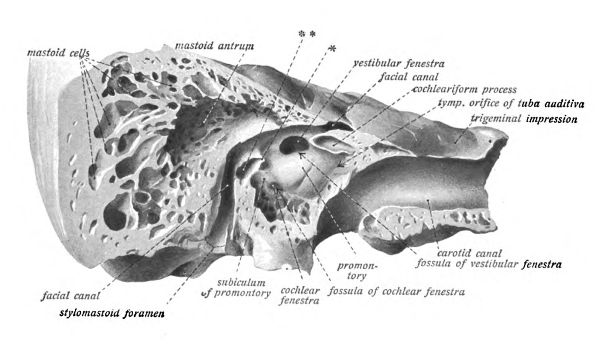
The right tympanic cavity: preparation of fig.791 after further removal of bone. English labels.
From 'Atlas and Textbook of Human Anatomy', 1911 (?), Vol. 3, fig.792, by Johannes Sobotta and J. Playfair McMurrich. Artist: K. Hajek. Retrieved from Sobotta's Anatomy plates at Wikimedia.
From 'Atlas and Textbook of Human Anatomy', 1911 (?), Vol. 3, fig.792, by Johannes Sobotta and J. Playfair McMurrich. Artist: K. Hajek. Retrieved from Sobotta's Anatomy plates at Wikimedia.
Anatomical structures in item:
Uploaded by: Student128
Netherlands, Leiden – Leiden University Medical Center, Leiden University
Cavitas tympani
Cellulae mastoideae
Antrum mastoideum
Fenestra vestibuli
Canalis nervi facialis
Processus cochleariformis
Canalis caroticus
Fossula fenestrae vestibuli
Promontorium tympani
Fossula fenestrae cochleae
Fenestra cochleae
Subiculum promontorii tympani
Foramen stylomastoideum
Creator(s)/credit: Prof.dr. Johannes Sobotta, anatomist
Requirements for usage
You are free to use this item.  Read more
Read more This item is in the Public Domain because its copyright has expired. You are not required to credit its creators when you use it. Nevertheless, it is adviced to do so. First, it is academically correct to pay tribute to the creators. Second, items of unknown origin might be classified as 'copyright infringement' by copyright controlling bodies, with possible resulting bills. Stating the item's source will prevent this. You can use the following text:
- For usage in print - copy and paste the line below:
- For digital usage (e.g. in PowerPoint, Impress, Word, Writer) - copy and paste the line below (optionally add the icon):
"Sobotta 1911 fig.792 - The right tympanic cavity - English labels" at AnatomyTOOL.org by Johannes Sobotta is in the Public Domain.
"Sobotta 1911 fig.792 - The right tympanic cavity - English labels" by Johannes Sobotta is in the Public Domain.


Comments