
nid: 58091
Additional formats:
None available
Description:
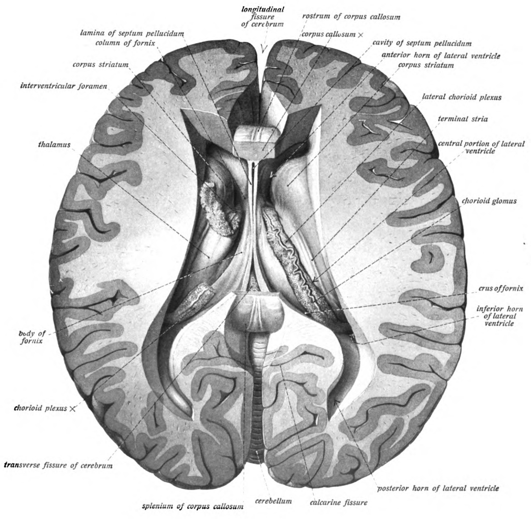
Lateral ventricles, fornix and septum pellucidum: transverse section showing tateral ventricles, fornix and septum pellucidum after partial removal of the corpus callosum. On the left side the choroid plexus has been divided. English labels.
From 'Atlas and Textbook of Human Anatomy', 1909, Vol. 3, fig.636, by Johannes Sobotta and J. Playfair McMurrich. Artist: K. Hajek. Retrieved from Sobotta's Anatomy plates at Wikimedia. Possible original source: Sobotta's atlas at Hathitrust Digital library.
From 'Atlas and Textbook of Human Anatomy', 1909, Vol. 3, fig.636, by Johannes Sobotta and J. Playfair McMurrich. Artist: K. Hajek. Retrieved from Sobotta's Anatomy plates at Wikimedia. Possible original source: Sobotta's atlas at Hathitrust Digital library.
Anatomical structures in item:
Uploaded by: Student128
Netherlands, Leiden – Leiden University Medical Center, Leiden University
Encephalon
Fornix
Pars centralis ventriculi lateralis
Septum pellucidum
Thalamus
Foramen interventriculare
Plexus choroideus
Sulcus calcarinus
Stria terminalis
Corpus striatum
Creator(s)/credit: Prof.dr. Johannes Sobotta, anatomist
Requirements for usage
You are free to use this item.  Read more
Read more This item is in the Public Domain because its copyright has expired. You are not required to credit its creators when you use it. Nevertheless, it is adviced to do so. First, it is academically correct to pay tribute to the creators. Second, items of unknown origin might be classified as 'copyright infringement' by copyright controlling bodies, with possible resulting bills. Stating the item's source will prevent this. You can use the following text:
- For usage in print - copy and paste the line below:
- For digital usage (e.g. in PowerPoint, Impress, Word, Writer) - copy and paste the line below (optionally add the icon):
"Sobotta 1909 fig.636 - Lateral ventricles, fornix and septum pellucidum, superior view - English labels" at AnatomyTOOL.org by Johannes Sobotta is in the Public Domain.


Comments