nid: 63491
Additional formats:
None available
Description:
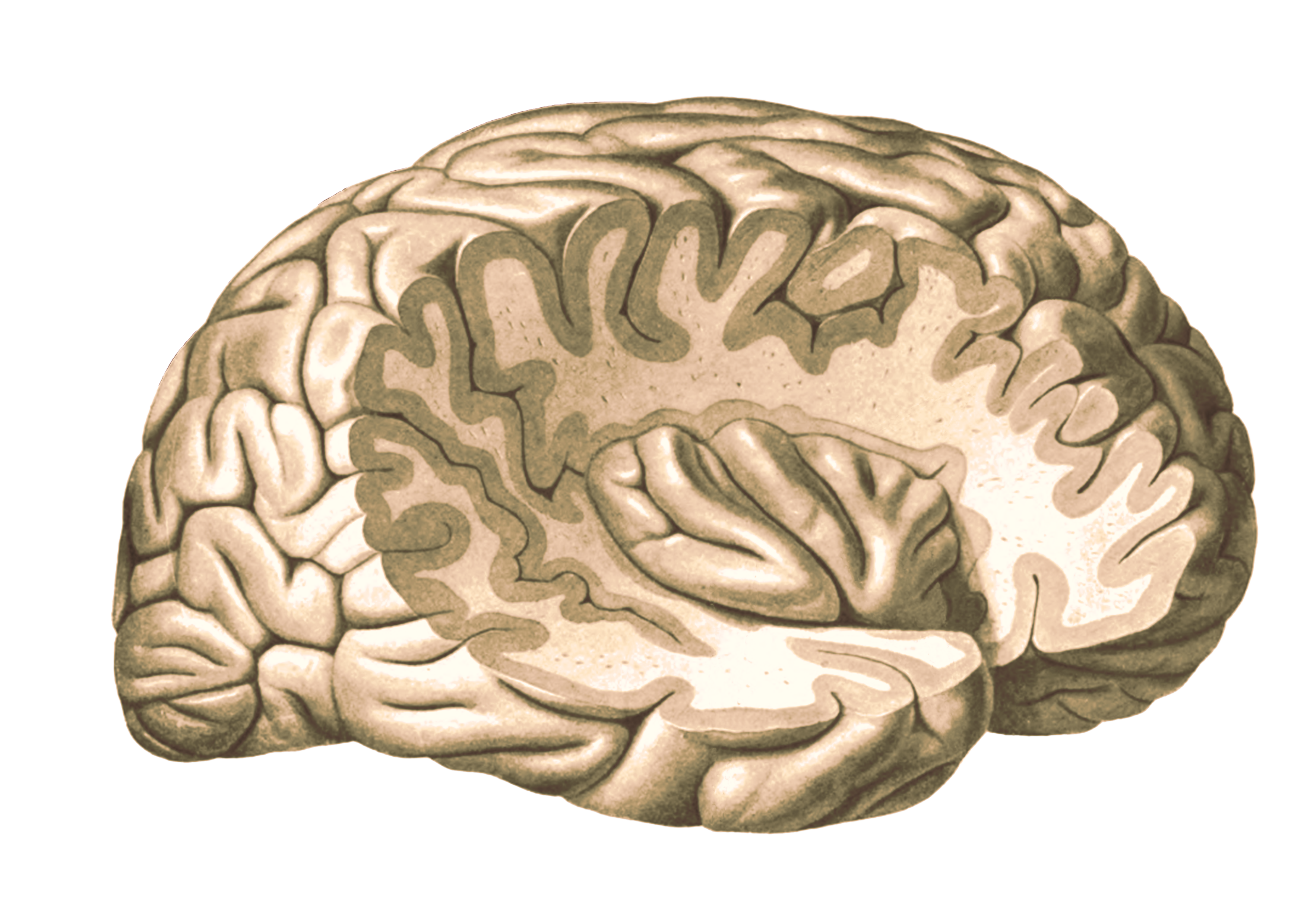
Cerebral cortex with insula: the insular convulsions have been exposed by removing portions of the frontal, temporal and parietal lobes. Coloured version without labels.
From 'Atlas and Textbook of Human Anatomy', 1909, Vol. 3, fig.633, by Johannes Sobotta and J. Playfair McMurrich. Artist: K. Hajek. Retrieved from Sobotta's Anatomy plates at Wikimedia. Possible original source: Sobotta's atlas at Hathitrust Digital library.
Coloured by Matty Spinder, MA, UMCU.
From 'Atlas and Textbook of Human Anatomy', 1909, Vol. 3, fig.633, by Johannes Sobotta and J. Playfair McMurrich. Artist: K. Hajek. Retrieved from Sobotta's Anatomy plates at Wikimedia. Possible original source: Sobotta's atlas at Hathitrust Digital library.
Coloured by Matty Spinder, MA, UMCU.
Anatomical structures in item:
Uploaded by: rva
Netherlands, Leiden – Leiden University Medical Center, Leiden University
Encephalon
Pallium
Insula
Gyrus longus insulae
Gyrus breves insulae
Lobus frontalis
Lobus temporalis
Lobus occipitalis
Lobus parietalis
Creator(s)/credit: Prof.dr. Johannes Sobotta, anatomist; Matty Spinder MD, MA, anatomist, image editing, UMC Utrecht
Requirements for usage
You are free to use this item if you follow the requirements of the license:  View license
 View license
 If you use this item you should credit it as follows:
- For usage in print - copy and paste the line below:
- For digital usage (e.g. in PowerPoint, Impress, Word, Writer) - copy and paste the line below (optionally add the license icon):
"Sobotta 1909 fig.633 - Cerebral cortex with insula - coloured, no labels" at AnatomyTOOL.org by Johannes Sobotta and Matty Spinder, UMC Utrecht, license: Creative Commons Attribution-ShareAlike
"Sobotta 1909 fig.633 - Cerebral cortex with insula - coloured, no labels" by Johannes Sobotta and Matty Spinder, UMC Utrecht, license: CC BY-SA


Comments