
nid: 62006
Additional formats:
None available
Description:
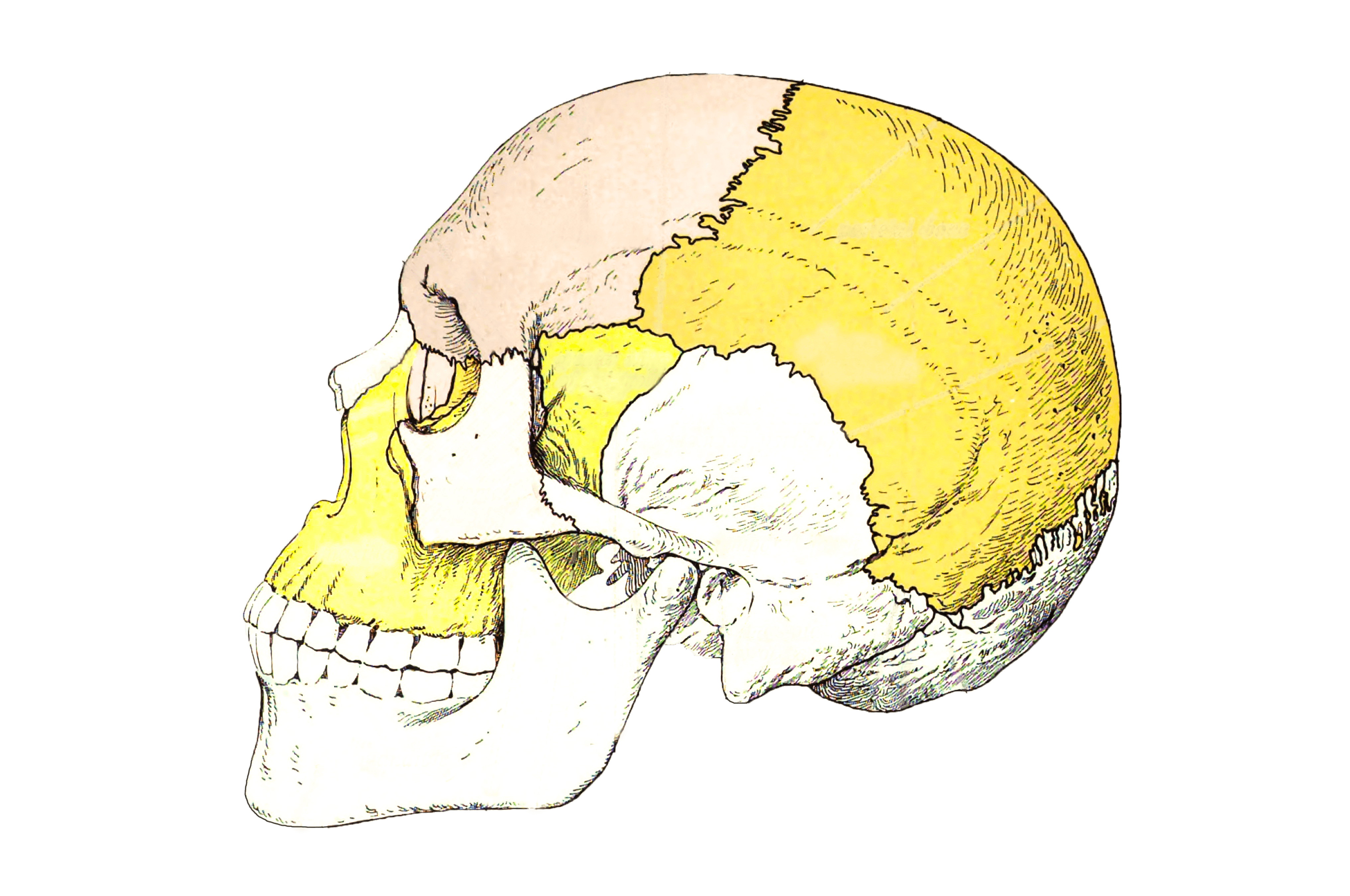
Bones of the skull, lateral view.
From 'Atlas and Textbook of Human Anatomy', 1909, Vol. 1, fig.39, by Johannes Sobotta and J. Playfair McMurrich. Artist: K. Hajek or A. Schmitson. Retrieved from Sobotta's Anatomy plates at Wikimedia. Possible original source: Sobotta's atlas at Hathitrust Digital library.
Image editing by dream_studio3.
Anatomical structures in item:
Uploaded by: rva
Netherlands, Leiden – Leiden University Medical Center, Leiden University
Cranium
Sutura sphenozygomatica
Os lacrimale
Sutura nasomaxillaris
Sutura lacrimomaxillaris
Processus frontalis maxillae
Os nasale
Spina nasalis anterior corporis maxillae
Maxilla
Os zygomaticum
Os frontale
Os parietale
Planum temporale
Os temporale
Pars squamosa ossis temporalis
Fossa temporalis
Processus mastoideus
Mandibula
Ala major ossis sphenoidalis
Os sphenoidale
Foramen mentale
Processus coronoideus mandibulae
Sutura sphenosquamosa
Arcus zygomaticus
Processus condylaris mandibulae
Meatus acusticus externus
Sutura parietomastoidea
Os occipitale
Squama occipitalis
Margo lambdoideus ossis occipitalis
Sutura squamosa
Inferior temporal line
Superior temporal line
Sutura coronalis
Sutura sphenoparietalis
Sutura sphenofrontalis
Creator(s)/credit: Prof Johannes Sobotta, Anatomist; dream_studio3 BA, image editing
Requirements for usage
You are free to use this item if you follow the requirements of the license:  View license
View license If you use this item you should credit it as follows:
- For usage in print - copy and paste the line below:
- For digital usage (e.g. in PowerPoint, Impress, Word, Writer) - copy and paste the line below (optionally add the license icon):
"Sobotta 1909 fig.39 - bones of the skull, lateral view - no labels" at AnatomyTOOL.org by Johannes Sobotta and dream_studio3, license: Creative Commons Attribution-ShareAlike
"Sobotta 1909 fig.39 - bones of the skull, lateral view - no labels" by Johannes Sobotta and dream_studio3, license: CC BY-SA


Comments