nid: 61629
Additional formats:
None available
Description:
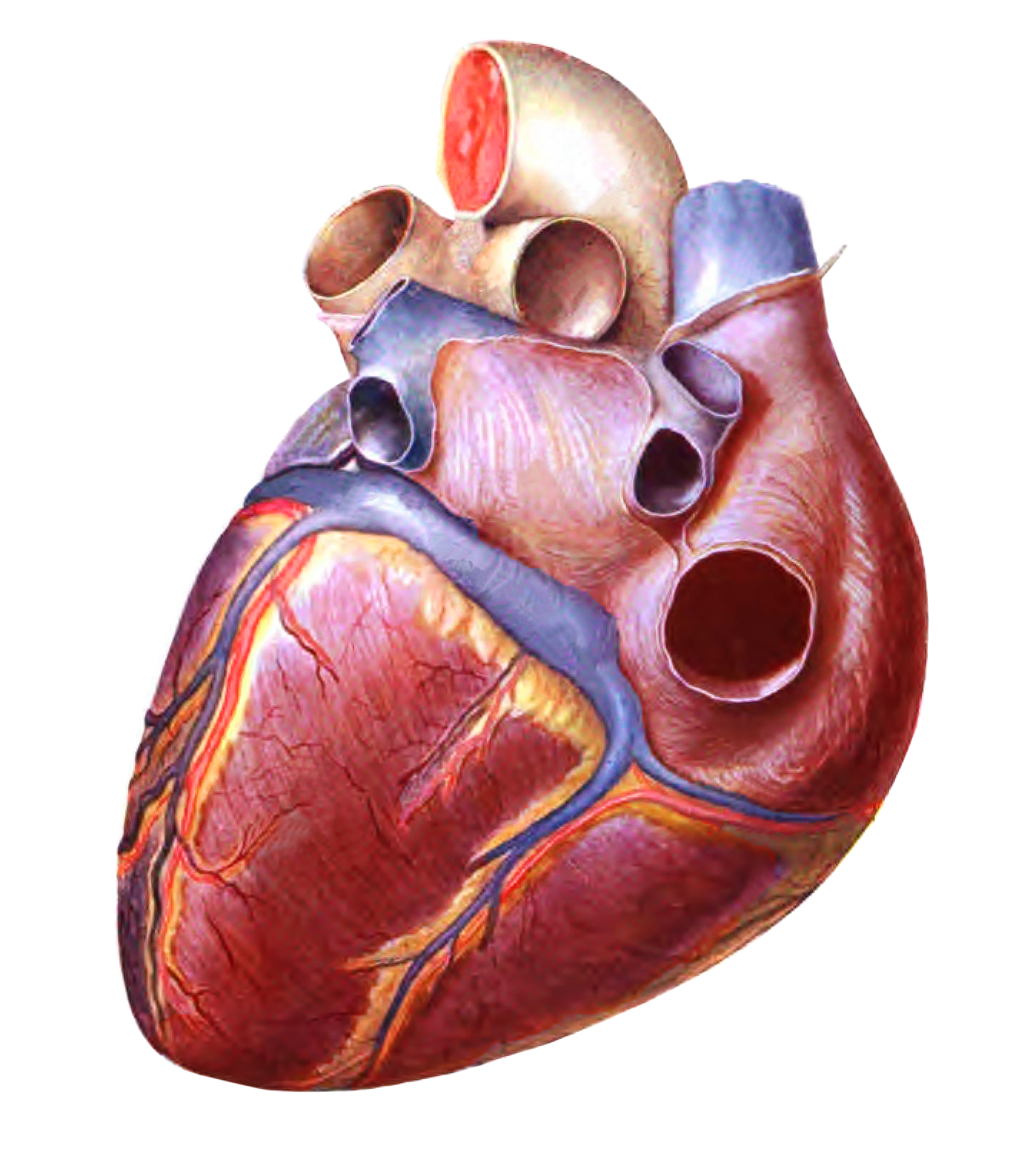
Diaphragmatic surface of the heart.
From 'Atlas and Textbook of Human Anatomy', 1906, Vol. 2, fig.519, by Johannes Sobotta and J. Playfair McMurrich. Artist: K. Hajek. Retrieved from Sobotta's Anatomy plates at Wikimedia. Possible original source: Sobotta's atlas at archive.org.
Image editing by dream_studio3.
From 'Atlas and Textbook of Human Anatomy', 1906, Vol. 2, fig.519, by Johannes Sobotta and J. Playfair McMurrich. Artist: K. Hajek. Retrieved from Sobotta's Anatomy plates at Wikimedia. Possible original source: Sobotta's atlas at archive.org.
Image editing by dream_studio3.
Anatomical structures in item:
Uploaded by: rva
Netherlands, Leiden – Leiden University Medical Center, Leiden University
Cor
Atrium sinistrum
Ventriculus sinister
Truncus pulmonalis
Arcus aortae
Vena cava inferior
Vena cava superior
Venae pulmonales
Sinus coronarius
Sulcus interventricularis posterior
Apex cordis
Auricula sinistra
Sulcus terminalis cordis
Creator(s)/credit: Prof Johannes Sobotta, Anatomist; dream_studio3 BA, image editing
Requirements for usage
You are free to use this item if you follow the requirements of the license:  View license
 View license
 If you use this item you should credit it as follows:
- For usage in print - copy and paste the line below:
- For digital usage (e.g. in PowerPoint, Impress, Word, Writer) - copy and paste the line below (optionally add the license icon):
"Sobotta 1906 fig.519 - diaphragmatic surface of the heart - no labels" at AnatomyTOOL.org by Johannes Sobotta and dream_studio3, license: Creative Commons Attribution-ShareAlike
"Sobotta 1906 fig.519 - diaphragmatic surface of the heart - no labels" by Johannes Sobotta and dream_studio3, license: CC BY-SA


Comments