nid: 61985
Additional formats:
None available
Description:
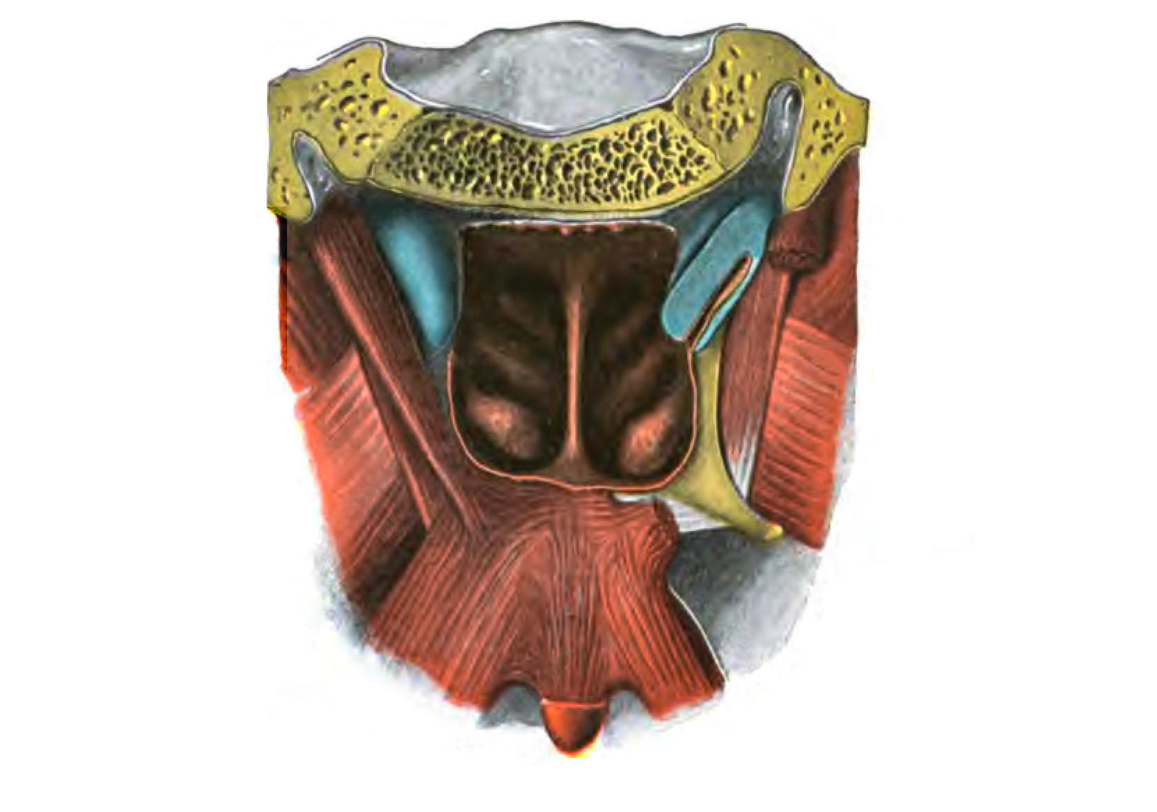
Palatine muscles: posterior view. On the right side the m. levator veli has been removed and the tuba auditiva is opened.
From 'Atlas and Textbook of Human Anatomy', 1906, Vol. 2, fig.329 , by Johannes Sobotta and J. Playfair McMurrich. Artist: K. Hajek. Retrieved from Sobotta's Anatomy plates at Wikimedia. Possible original source: Sobotta's atlas at archive.org.
Image editing by Theo Photographyx
From 'Atlas and Textbook of Human Anatomy', 1906, Vol. 2, fig.329 , by Johannes Sobotta and J. Playfair McMurrich. Artist: K. Hajek. Retrieved from Sobotta's Anatomy plates at Wikimedia. Possible original source: Sobotta's atlas at archive.org.
Image editing by Theo Photographyx
Anatomical structures in item:
Uploaded by: rva
Netherlands, Leiden – Leiden University Medical Center, Leiden University
Palatum
Musculus levator veli palatini
Musculus tensor veli palatini
Musculus palatopharyngeus
Hamulus pterygoideus
Tuba auditoria (auditiva)
Concha nasalis superior
Concha nasalis media
Concha nasalis inferior
Creator(s)/credit: Prof.dr. Johannes Sobotta, anatomist; Theo Photographyx, photographer, image editing
Requirements for usage
You are free to use this item if you follow the requirements of the license:  View license
 View license
 If you use this item you should credit it as follows:
- For usage in print - copy and paste the line below:
- For digital usage (e.g. in PowerPoint, Impress, Word, Writer) - copy and paste the line below (optionally add the license icon):
"Sobotta 1906 fig.329 - Palatine muscles - no labels" at AnatomyTOOL.org by Johannes Sobotta and Theo Photographyx, license: Creative Commons Attribution-ShareAlike


Comments