
nid: 62961
Additional formats:
None available
Description:
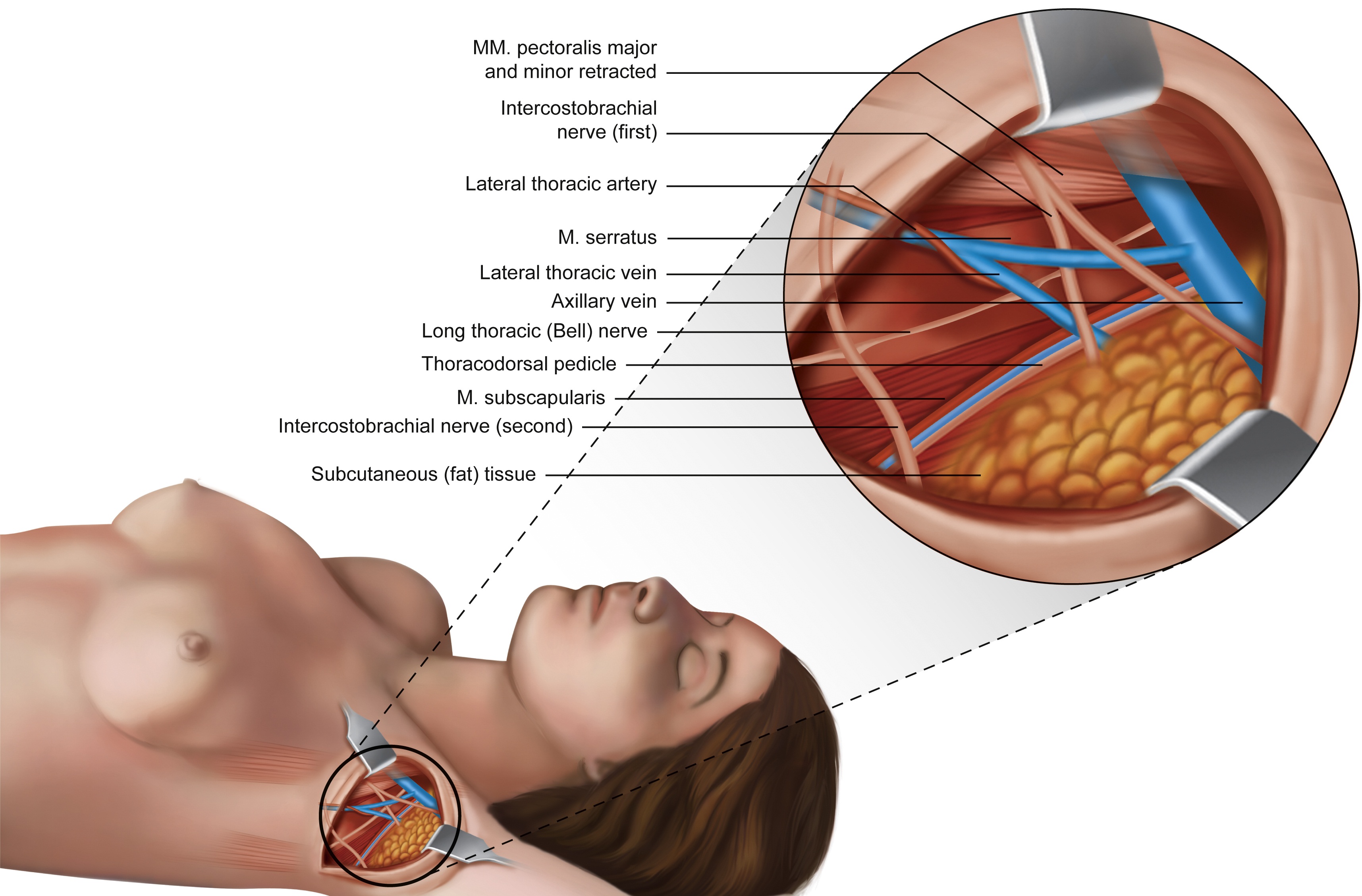
Axilla after axillary lymph node dissection. The first and second intercostobrachial nerve are visible. This paper showed that in about 40% of the cases divisions of the ICBN were observed in the central region of the axilla. English labels.
The image and part of the description is retrieved from the paper: Soares EW. Anatomical variations of the axilla. Springerplus. 2014 Jun 24;3:306, published under a CC BY license.
The image and part of the description is retrieved from the paper: Soares EW. Anatomical variations of the axilla. Springerplus. 2014 Jun 24;3:306, published under a CC BY license.
Anatomical structures in item:
Uploaded by: rva
Netherlands, Leiden – Leiden University Medical Center, Leiden University
Nodi lymphoidei axillares
Regio axillaris
Musculus pectoralis major
Musculus pectoralis minor
Nervi intercostobrachiales
Arteria thoracica lateralis
Musculus serratus anterior
Vena thoracica lateralis
Vena axillaris
Nervus thoracicus longus
Nervus thoracodorsalis
Musculus subscapularis
Panniculus adiposus (Tela subcutanea)
Creator(s)/credit: Emerson Wander Silva Soares PhD, MD, surgeon
Requirements for usage
You are free to use this item if you follow the requirements of the license:  View license
View license If you use this item you should credit it as follows:
- For usage in print - copy and paste the line below:
- For digital usage (e.g. in PowerPoint, Impress, Word, Writer) - copy and paste the line below (optionally add the license icon):
"Soares - Drawing Axilla after axillary lymph node dissection - English labels" at AnatomyTOOL.org by Emerson Wander Silva Soares, license: Creative Commons Attribution


Comments