
nid: 62236
Additional formats:
None available
Description:
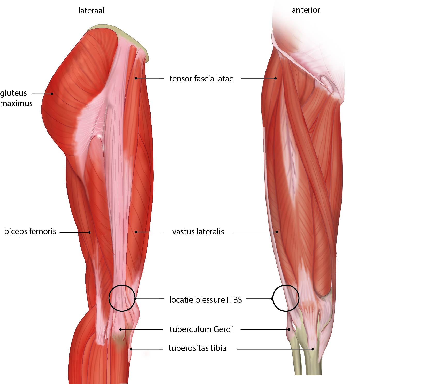
Lateral and anterior view of muscles of thigh and location Iliotibial band syndrome. Iliotibial band syndrome (ITBS) can occur due to friction between the iliotibial band and the lateral epicondyle of the femur. ITBS is a type of overuse injury known as a Runner's knee. Dutch labels
Anatomical structures in item:
Uploaded by: rva
Netherlands, Leiden – Leiden University Medical Center, Leiden University
Musculus gluteus maximus
Musculus tensor fasciae latae
Femur
Musculus biceps femoris
Musculus vastus lateralis
Tractus iliotibialis
Tuberositas tibiae
Musculus vastus medialis
Musculus quadriceps femoris
Musculus rectus femoris
Musculus pectineus
Musculus sartorius
Musculus gracilis
Musculus adductor longus
Creator(s)/credit: Ron Slagter NZIMBI, medical illustrator
Requirements for usage
You are free to use this item if you follow the requirements of the license:  View license
View license If you use this item you should credit it as follows:
- For usage in print - copy and paste the line below:
- For digital usage (e.g. in PowerPoint, Impress, Word, Writer) - copy and paste the line below (optionally add the license icon):
"Slagter - Drawing Lateral and anterior view of muscles of thigh and location Iliotibial band syndrome - Dutch labels" at AnatomyTOOL.org by Ron Slagter, license: Creative Commons Attribution-NonCommercial-ShareAlike


Comments