
nid: 63209
Additional formats:
None available
Description:
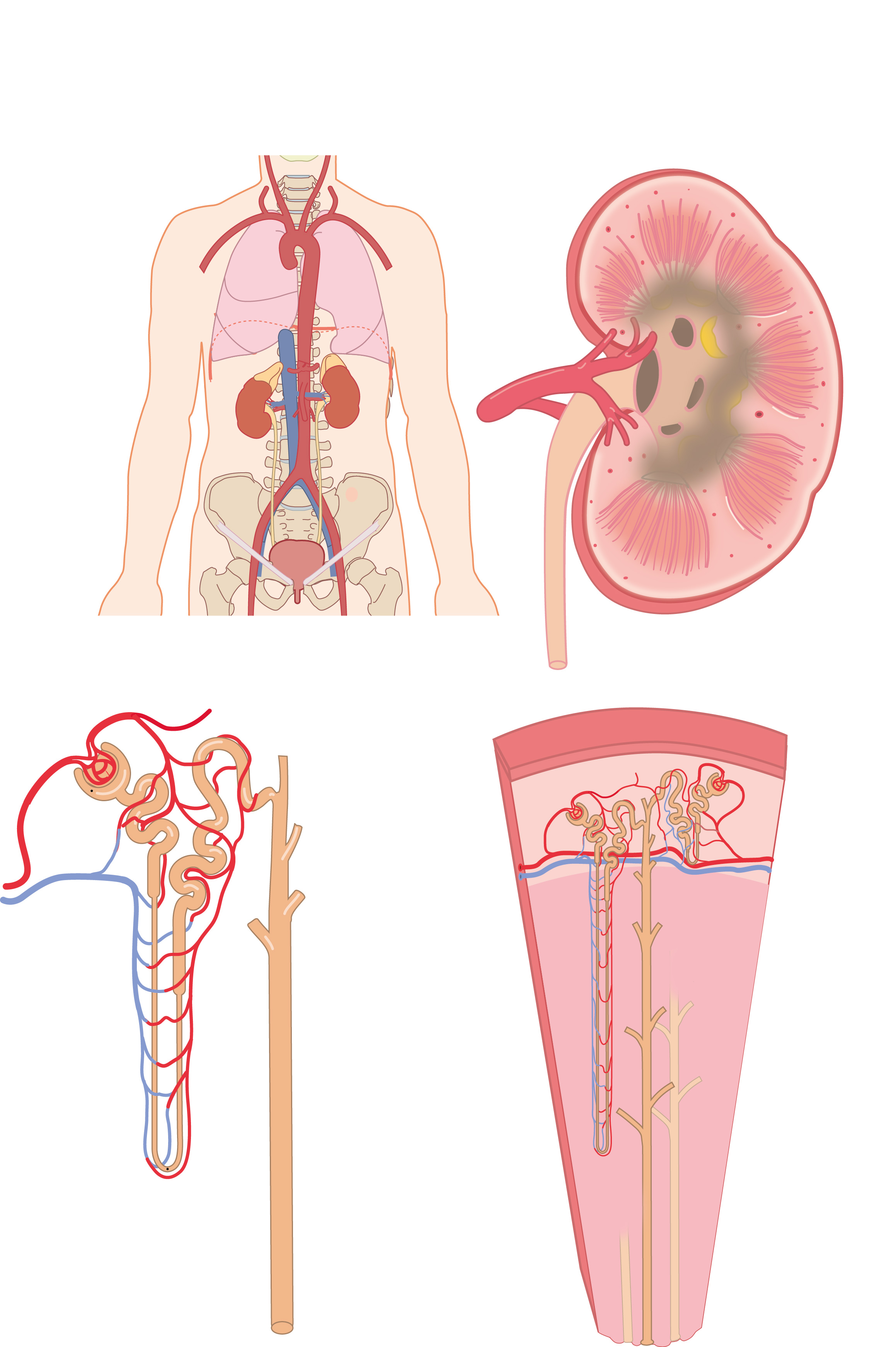
Kidney macroscopic and microscopic anatomy. Four drawings show the anatomy of the kidney, these drawings depict (clockwise): position in the body, cross-section of the kidney, anatomy of the medulla, and the anatomy of a juxtamedullary nephron. Version without labels
Anatomical structures in item:
Uploaded by: rva
Netherlands, Leiden – Leiden University Medical Center, Leiden University
Glandula suprarenalis
Ren (Nephros)
Ureter
Vesica urinaria
Urethra
Medulla renalis
Pelvis renalis
Capsula glomerularis
Tubuli seminiferi contorti
Tubuli seminiferi recti
Creator(s)/credit: Ron Slagter NZIMBI, medical illustrator
Requirements for usage
You are free to use this item if you follow the requirements of the license:  View license
View license If you use this item you should credit it as follows:
- For usage in print - copy and paste the line below:
- For digital usage (e.g. in PowerPoint, Impress, Word, Writer) - copy and paste the line below (optionally add the license icon):
"Slagter - Drawing Kidney macroscopic and microscopic anatomy - no labels" at AnatomyTOOL.org by Ron Slagter, license: Creative Commons Attribution-NonCommercial-ShareAlike


Comments