
nid: 59163
Additional formats:
None available
Description:
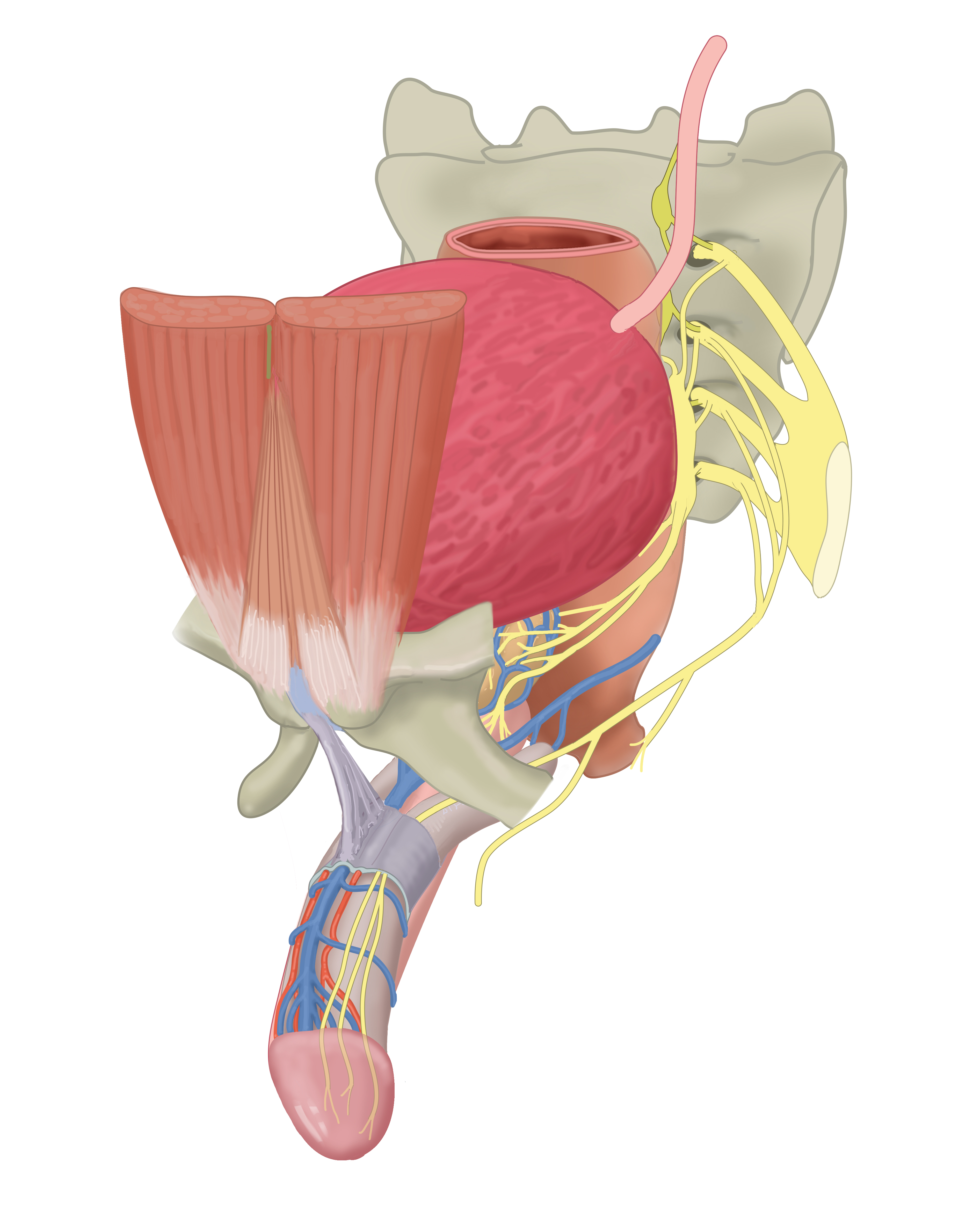
Rectus muscle, pyramidalis muscle and suspensory ligament of the penis. The small bilateral pyramidalis muscle lies anterior to the rectus muscle. The anterior rectus sheath (not shown here) is continuous with the suspensory ligament of the penis. The suspensory ligament attaches to root of the penis to the pubic symphysis. No labels.
Illustration by Ron Slagter and Marco DeRuiter for course 'Surgical Anatomy of the lesser pelvis' by the 'Urologisch Opleidings Instituut', the Netherlands.
Illustration by Ron Slagter and Marco DeRuiter for course 'Surgical Anatomy of the lesser pelvis' by the 'Urologisch Opleidings Instituut', the Netherlands.
Anatomical structures in item:
Uploaded by: Siem Zethof
Netherlands, Leiden – Leiden University Medical Center, Leiden University
Musculus pyramidalis
Ligamentum suspensorium penis
Creator(s)/credit: Ron Slagter NZIMBI, medical illustrator, LUMC; Prof. Marco DeRuiter PhD, anatomist, LUMC
Requirements for usage
You are free to use this item if you follow the requirements of the license:  View license
View license If you use this item you should credit it as follows:
- For usage in print - copy and paste the line below:
- For digital usage (e.g. in PowerPoint, Impress, Word, Writer) - copy and paste the line below (optionally add the license icon):
"Slagter - Drawing Innervation and ligaments of the penis - no labels" at AnatomyTOOL.org by Ron Slagter, LUMC and Marco DeRuiter, LUMC, license: Creative Commons Attribution-NonCommercial-ShareAlike
"Slagter - Drawing Innervation and ligaments of the penis - no labels" by Ron Slagter, LUMC and Marco DeRuiter, LUMC, license: CC BY-NC-SA


Comments