
nid: 62104
Additional formats:
None available
Description:
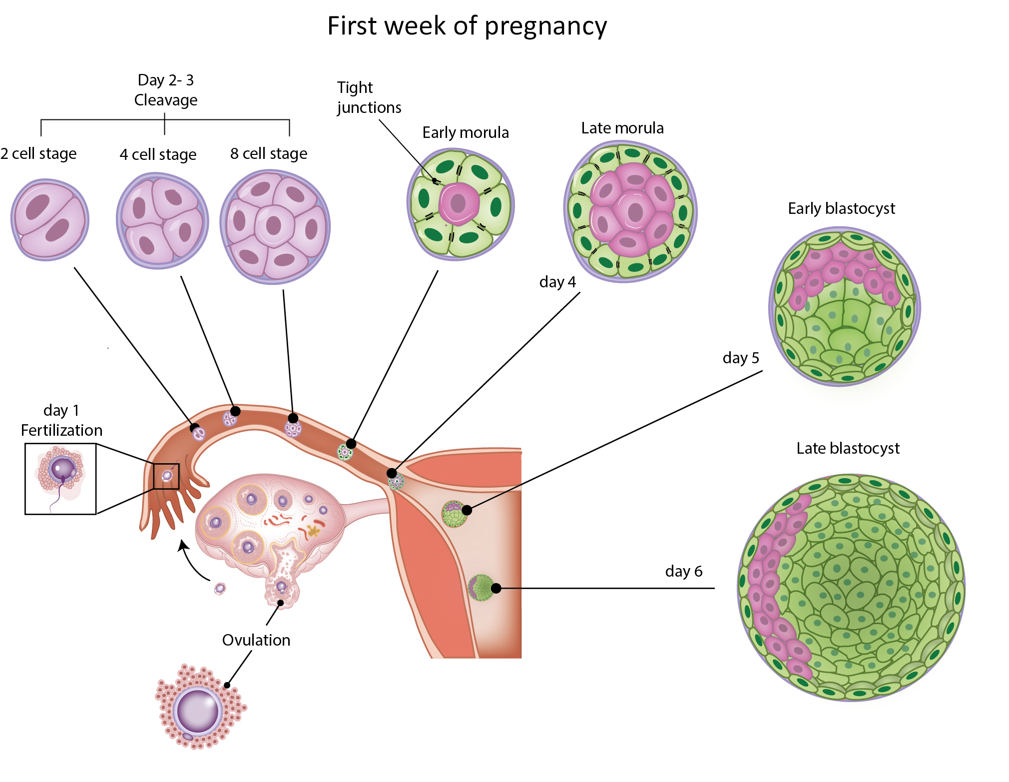
First week of pregnancy. After fertilisation, mitotic division of the zygote occurs. After 4 divisions, the zygote reaches the morula stage (16-32 cells). This morula consists of an inner and outer cell mass and will become hollow to become a blastocyst. English labels. This drawing belongs to a series of drawings of early embryology.
Anatomical structures in item:
Uploaded by: rva
Netherlands, Leiden – Leiden University Medical Center, Leiden University
Ovarium
Tuba uterina (Salpinx)
Uterus
Creator(s)/credit: Ron Slagter NZIMBI, medical illustrator; Hope Wicks MBSc, medical student, LUMC; drs Cindy J.M. Hulsman, PhD student, MUMC+; Jill P.J.M. Hikspoors PhD, assistant professor of anatomy, MUMC+; O. Paul Gobée MD, anatomist, LUMC
Requirements for usage
You are free to use this item if you follow the requirements of the license:  View license
View license If you use this item you should credit it as follows:
- For usage in print - copy and paste the line below:
- For digital usage (e.g. in PowerPoint, Impress, Word, Writer) - copy and paste the line below (optionally add the license icon):
"Slagter - Drawing First week of pregnancy - English labels" at AnatomyTOOL.org by Ron Slagter, Hope Wicks, LUMC, Cindy J.M. Hulsman, MUMC+ et al, license: Creative Commons Attribution-NonCommercial-ShareAlike
"Slagter - Drawing First week of pregnancy - English labels" by Ron Slagter, Hope Wicks, LUMC, Cindy J.M. Hulsman, MUMC+ et al, license: CC BY-NC-SA


Comments