
nid: 62362
Additional formats:
None available
Description:
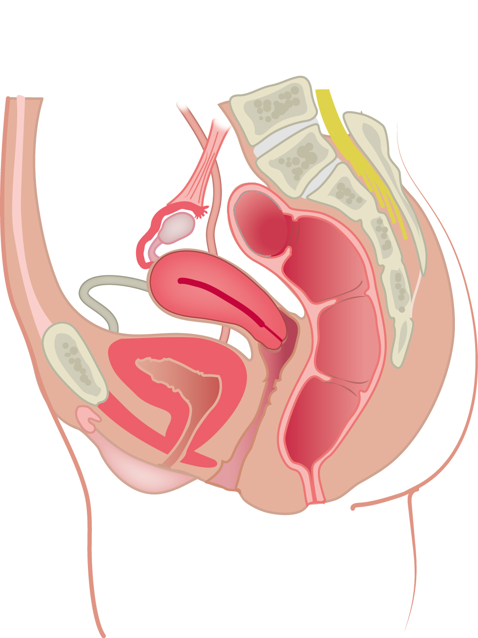
Female pelvis in sagittal section. The position of the female urogenital organs in the pelvis is drawn in this image.
Anatomical structures in item:
Uploaded by: rva
Netherlands, Leiden – Leiden University Medical Center, Leiden University
Pelvis
Vesica urinaria
Ureter
Anus
Rectum
Os sacrum [vertebrae sacrales I - V]
Vagina
Uterus
Pubis
Ovarium
Tuba uterina (Salpinx)
Ligamentum suspensorium ovarii
Creator(s)/credit: Ron Slagter NZIMBI, medical illustrator
Requirements for usage
You are free to use this item if you follow the requirements of the license:  View license
View license If you use this item you should credit it as follows:
- For usage in print - copy and paste the line below:
- For digital usage (e.g. in PowerPoint, Impress, Word, Writer) - copy and paste the line below (optionally add the license icon):
"Slagter - Drawing Female pelvis in sagittal section - no labels" at AnatomyTOOL.org by Ron Slagter, license: Creative Commons Attribution-NonCommercial-ShareAlike


Comments