nid: 62268
Additional formats:
None available
Description:
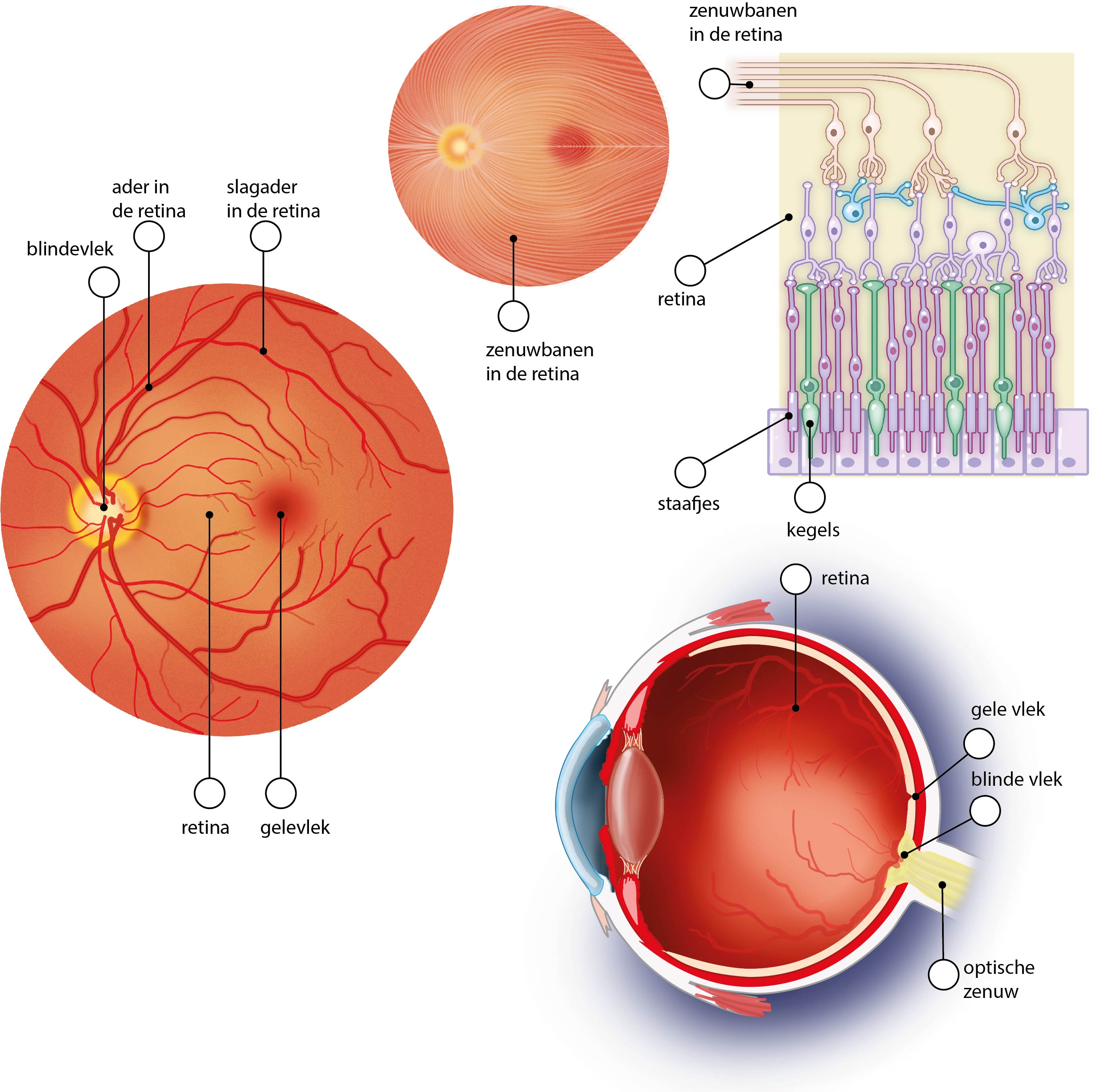
Eye: retina and retinal nerve fiber layer. Clockwise, starting left: the first and second drawing show the anatomy of the retina, the third drawing shows the retinal nerve fiber layer with rods and cones, and the fourth drawing shows the eye in a sagittal section. Dutch labels.
Anatomical structures in item:
Uploaded by: rva
Netherlands, Leiden – Leiden University Medical Center, Leiden University
Stratum segmentorum externorum et internorum retinae
Pars caeca retinae
Arteria centralis retinae
Vena centralis retinae
Macula lutea
Retina
Bulbus oculi
Oculus et structurae pertinentes
Oculus
Stratum neurofibrarum retinae
Nervus opticus
Creator(s)/credit: Ron Slagter NZIMBI, medical illustrator
Requirements for usage
You are free to use this item if you follow the requirements of the license:  View license
 View license
 If you use this item you should credit it as follows:
- For usage in print - copy and paste the line below:
- For digital usage (e.g. in PowerPoint, Impress, Word, Writer) - copy and paste the line below (optionally add the license icon):
"Slagter - Drawing Eye: retina and retinal nerve fiber layer - Dutch labels" at AnatomyTOOL.org by Ron Slagter, license: Creative Commons Attribution-NonCommercial-ShareAlike


Comments