
nid: 60724
Additional formats:
None available
Description:
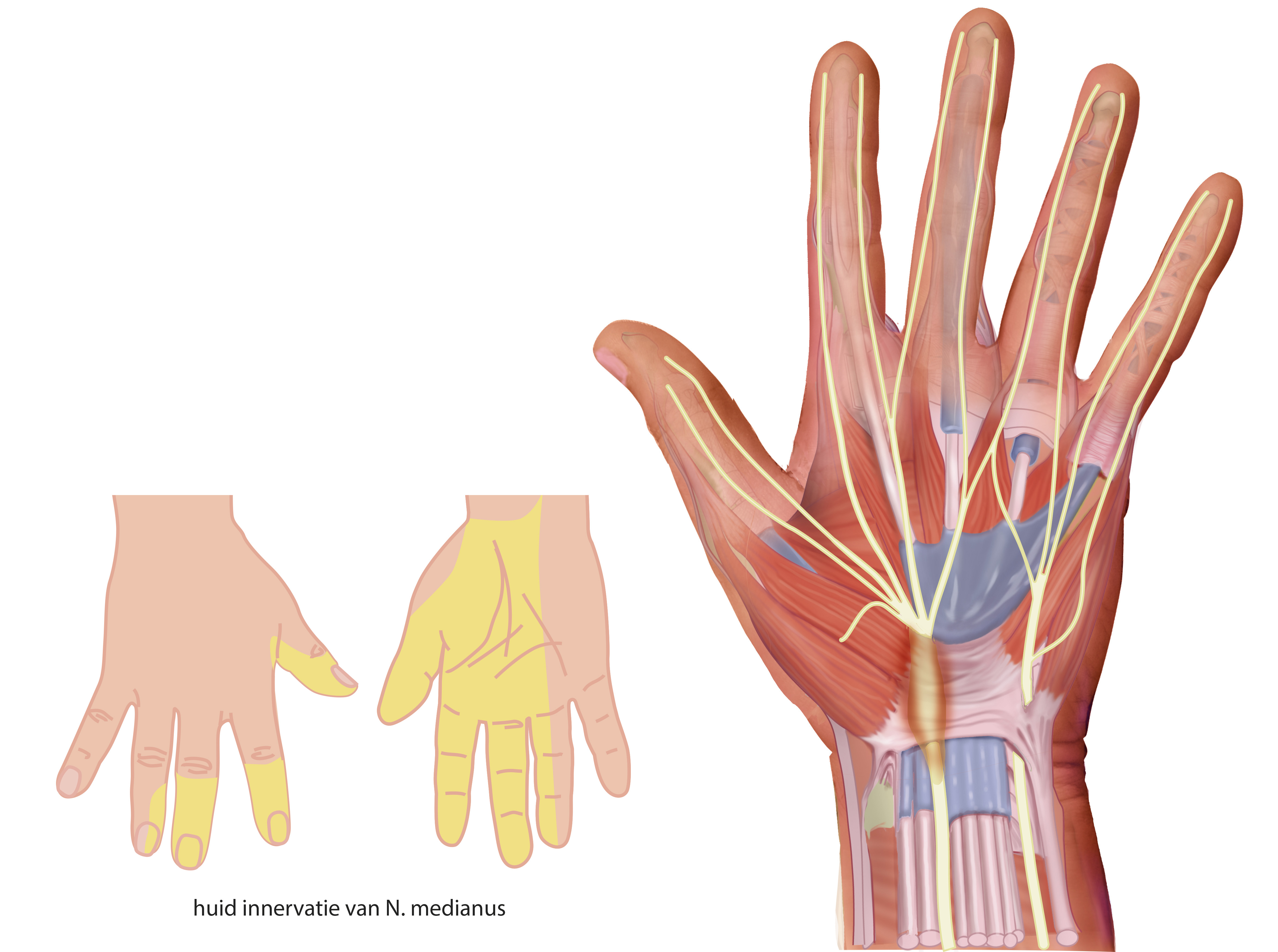
The image shows where the median nerve is compressed by the transverse carpal ligament (flexor retinaculum). Also it shows the skin innervation distribution area of the median nerve.
Illustration by Ron Slagter.
Illustration by Ron Slagter.
Anatomical structures in item:
Uploaded by: opgobee
Netherlands, Leiden – Leiden University Medical Center, Leiden University
Manus
Nervus medianus
Retinaculum musculorum flexorum manus
Creator(s)/credit: Ron Slagter NZIMBI, medical illustrator
Requirements for usage
You are free to use this item if you follow the requirements of the license:  View license
View license If you use this item you should credit it as follows:
- For usage in print - copy and paste the line below:
- For digital usage (e.g. in PowerPoint, Impress, Word, Writer) - copy and paste the line below (optionally add the license icon):
"Slagter - Drawing Carpal tunnel syndrome - Dutch labels" at AnatomyTOOL.org by Ron Slagter, license: Creative Commons Attribution-NonCommercial-ShareAlike


Comments