
nid: 62361
Additional formats:
None available
Description:
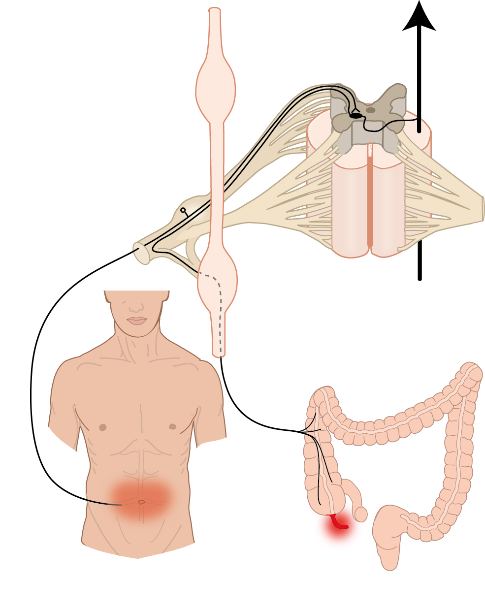
Inflamed appendix and pain route. This image depicts the anatomical background of the classical pain presentation of (early) appendicitis. The pain signal from the appendix follows the indicated route to arrive at a certain level of the spinal cord. Sensory information from the abdominal wall in the umbilical area arrives at the same level in the spinal cord. The origin of the pain signal then cannot be distinguished anymore, leading to so called 'referred pain' experienced in the abdominal wall around the umbilicus.
Anatomical structures in item:
Uploaded by: rva
Netherlands, Leiden – Leiden University Medical Center, Leiden University
Appendix vermiformis
Vena dorsalis
Ganglion sensorium nervi spinalis
Radix posterior (Nervus spinalis)
Radix anterior (Nervus spinalis)
Medulla spinalis
Truncus sympathicus
Ganglion sympathicum
Creator(s)/credit: Ron Slagter NZIMBI, medical illustrator; O. Paul Gobée MD, anatomist, LUMC
Requirements for usage
You are free to use this item if you follow the requirements of the license:  View license
View license If you use this item you should credit it as follows:
- For usage in print - copy and paste the line below:
- For digital usage (e.g. in PowerPoint, Impress, Word, Writer) - copy and paste the line below (optionally add the license icon):
"Slagter - Drawing appendicitis, pain route and referred pain - no labels" at AnatomyTOOL.org by Ron Slagter and O. Paul Gobée, LUMC, license: Creative Commons Attribution-NonCommercial-ShareAlike


Comments