nid: 63241
Additional formats:
None available
Description:
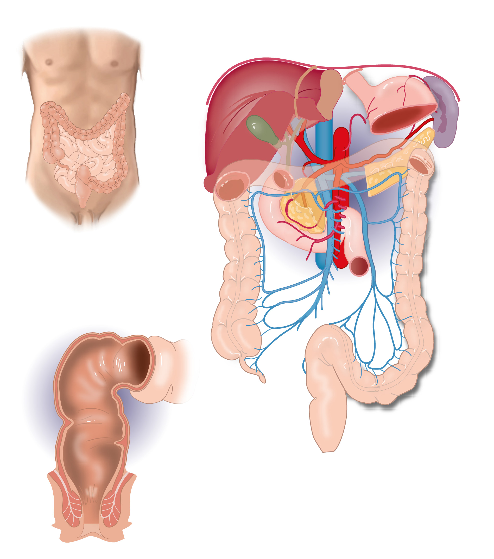
Anatomy of the abdominal gastrointestinal tract. The upper left drawing is a overview of the colon, ileum and jejunum; the lower left drawing is a detailed drawing of the rectum; and the right drawing is an overview of the organs of the gastrointestinal tract and their blood vessels. Version without labels.
Erratum:
16 March 2025 On the right drawing, the splenic vein incorrectly is drawn posterior of sup. mesent. art. but should run anterior of SMA. Thanks to Tijmen Vermeulen, medical student LUMC who noticed this error. Corrected version: see https://anatomytool.org/content/slagter-drawing-portal-vein-no-labels
Anatomical structures in item:
Uploaded by: rva
Netherlands, Leiden – Leiden University Medical Center, Leiden University
Abdomen
Systema digestorium
Colon
Ileum
Hepar
Vesica biliaris (Fellea)
Vena portae hepatis
Arteria mesenterica superior
Duodenum
Colon ascendens
Vena cava inferior
Aorta abdominalis
Colon sigmoideum
Colon descendens
Vena mesenterica inferior
Vena mesenterica superior
Pancreas
Cauda pancreatis
Colon transversum
Splen
Ventriculus
Ductus biliaris
Truncus coeliacus
Arteria lienalis
Arteria hepatica communis
Arteria hepatica propria
Arteria gastroduodenalis
Rectum
Musculus levator ani
Musculus sphincter ani externus
Musculus sphincter ani internus
Creator(s)/credit: Ron Slagter NZIMBI, medical illustrator
Requirements for usage
You are free to use this item if you follow the requirements of the license:  View license
 View license
 If you use this item you should credit it as follows:
- For usage in print - copy and paste the line below:
- For digital usage (e.g. in PowerPoint, Impress, Word, Writer) - copy and paste the line below (optionally add the license icon):
"Slagter - Drawing Anatomy of the abdominal gastrointestinal tract - no labels" at AnatomyTOOL.org by Ron Slagter, license: Creative Commons Attribution-NonCommercial-ShareAlike


Comments