
nid: 62613
Additional formats:
None available
Description:
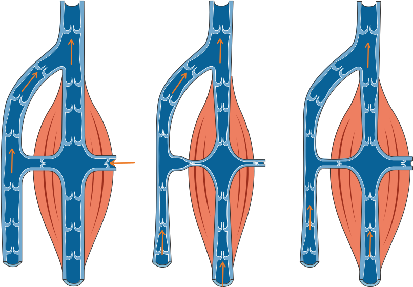
Venous blood flow due to muscle contraction. Some veins are located in between muscles (e.g. in the lower extremity). When the muscles contract the pressure increases, the valves will open so that blood will flow towards the heart. For similar images and pathology, see smart.servier.com.
Anatomical structures in item:
Uploaded by: rva
Netherlands, Leiden – Leiden University Medical Center, Leiden University
Vena
Valvula venosa
Creator(s)/credit: Servier Medical Art
Requirements for usage
You are free to use this item if you follow the requirements of the license:  View license
View license If you use this item you should credit it as follows:
- For usage in print - copy and paste the line below:
- For digital usage (e.g. in PowerPoint, Impress, Word, Writer) - copy and paste the line below (optionally add the license icon):
"Servier - Drawing Venous blood flow due to muscle contraction - no labels" at AnatomyTOOL.org by Servier Medical Art, license: Creative Commons Attribution


Comments