
nid: 61939
Additional formats:
None available
Description:
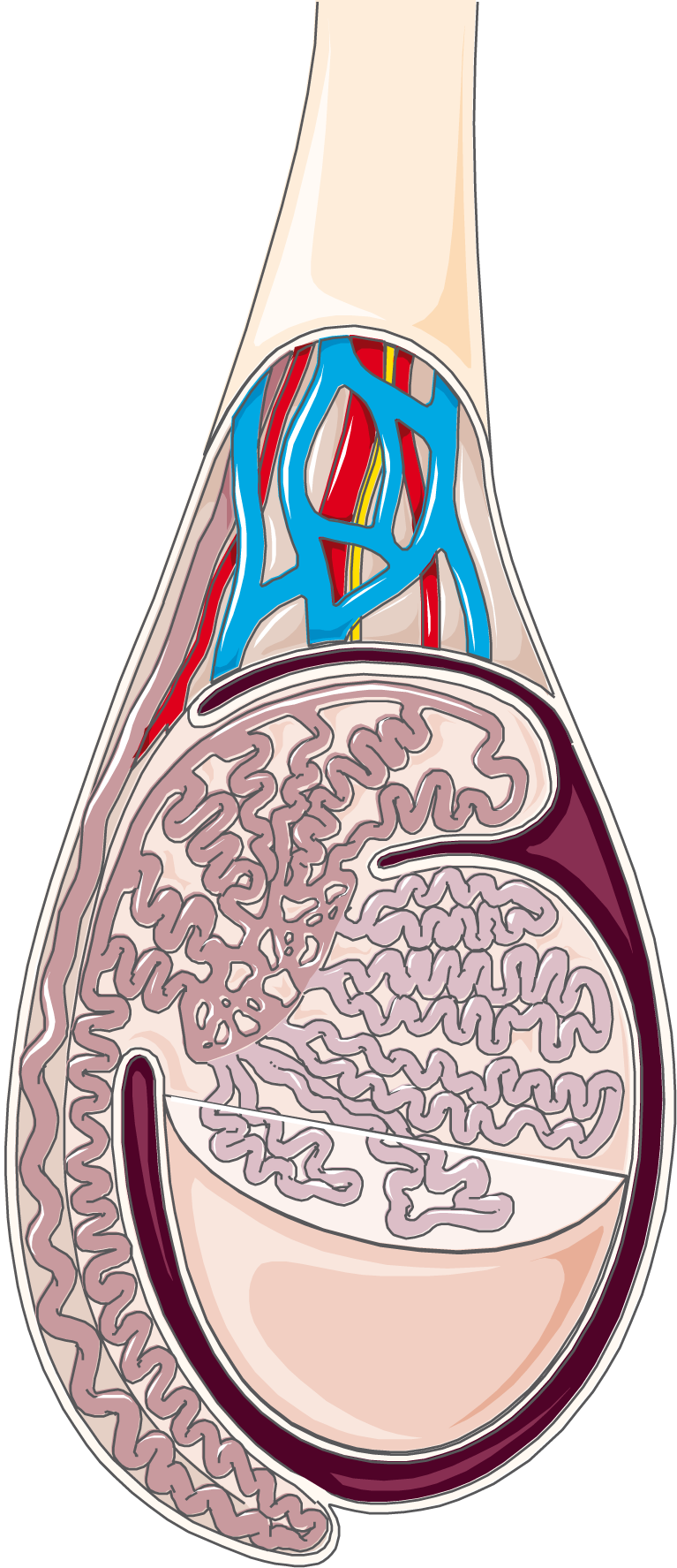
Testis and epididymis. For similar images and pathology, see smart.servier.com.
Anatomical structures in item:
Uploaded by: rva
Netherlands, Leiden – Leiden University Medical Center, Leiden University
Epididymis
Testis
Caput epididymidis
Ductuli efferentes testis
Arteria testicularis
Plexus pampiniformis
Parietal layer of tunica vaginalis of testis
Ductus deferens
Tunica albuginea testis
Cauda epididymidis
Visceral layer of tunica vaginalis of testis
Tubuli seminiferi recti
Rete testis
Tubuli seminiferi contorti
Creator(s)/credit: Servier Medical Art
Requirements for usage
You are free to use this item if you follow the requirements of the license:  View license
View license If you use this item you should credit it as follows:
- For usage in print - copy and paste the line below:
- For digital usage (e.g. in PowerPoint, Impress, Word, Writer) - copy and paste the line below (optionally add the license icon):
"Servier - Drawing Testis and epididymis - no labels" at AnatomyTOOL.org by Servier Medical Art, license: Creative Commons Attribution


Comments