
nid: 62747
Additional formats:
None available
Description:
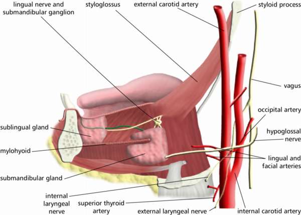
Submandibular structures: lateral view. The sublingual gland and submandibular gland are drawn, as well as their arteries and nerves. English labels.
This image by the Royal College of Surgeons of Ireland (RCSI) is retrieved from Health Education Assets Library (HEAL) of the University of Utah.
This image by the Royal College of Surgeons of Ireland (RCSI) is retrieved from Health Education Assets Library (HEAL) of the University of Utah.
Anatomical structures in item:
Uploaded by: rva
Netherlands, Leiden – Leiden University Medical Center, Leiden University
Nervus lingualis
Ganglion submandibulare
Musculus styloglossus
Arteria carotis externa
Processus styloideus
Nervus vagus
Arteria occipitalis
Nervus hypoglossus [XII]
Arteria lingualis
Arteria facialis
Arteria carotis interna
Ramus externus nervus laryngei superioris
Arteria thyroidea superior
Ramus internus nervus laryngei superioris
Glandula submandibularis
Musculus mylohyoideus
Glandula sublingualis
Creator(s)/credit: Royal College of Surgeons of Ireland
Requirements for usage
You are free to use this item if you follow the requirements of the license:  View license
View license If you use this item you should credit it as follows:
- For usage in print - copy and paste the line below:
- For digital usage (e.g. in PowerPoint, Impress, Word, Writer) - copy and paste the line below (optionally add the license icon):
"RCSI - Drawing Submandibular structures: lateral view - English labels" at AnatomyTOOL.org by Royal College of Surgeons of Ireland, license: Creative Commons Attribution-NonCommercial-ShareAlike


Comments