
nid: 62681
Additional formats:
None available
Description:
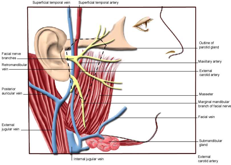
Parotid region. Muscles, nerves and arteries of the parotid region are drawn. The parotid gland itself is outlined. English labels.
This image by the Royal College of Surgeons of Ireland (RCSI) is retrieved from Health Education Assets Library (HEAL) of the University of Utah.
This image by the Royal College of Surgeons of Ireland (RCSI) is retrieved from Health Education Assets Library (HEAL) of the University of Utah.
Anatomical structures in item:
Uploaded by: rva
Netherlands, Leiden – Leiden University Medical Center, Leiden University
Regio parotideomasseterica
Venae temporales superficiales
Arteria temporalis superficialis
Nervus facialis [VII]
Vena retromandibularis
Vena auricularis posterior
Vena jugularis externa
Vena jugularis interna
Glandula submandibularis
Vena facialis
Ramus marginalis mandibularis nervus facialis
Musculus masseter
Arteria carotis externa
Arteria maxillaris
Glandula parotidea
Creator(s)/credit: Royal College of Surgeons of Ireland
Requirements for usage
You are free to use this item if you follow the requirements of the license:  View license
View license If you use this item you should credit it as follows:
- For usage in print - copy and paste the line below:
- For digital usage (e.g. in PowerPoint, Impress, Word, Writer) - copy and paste the line below (optionally add the license icon):
"RCSI - Drawing Parotid region - English labels" at AnatomyTOOL.org by Royal College of Surgeons of Ireland, license: Creative Commons Attribution-NonCommercial-ShareAlike


Comments