nid: 62735
Additional formats:
None available
Description:
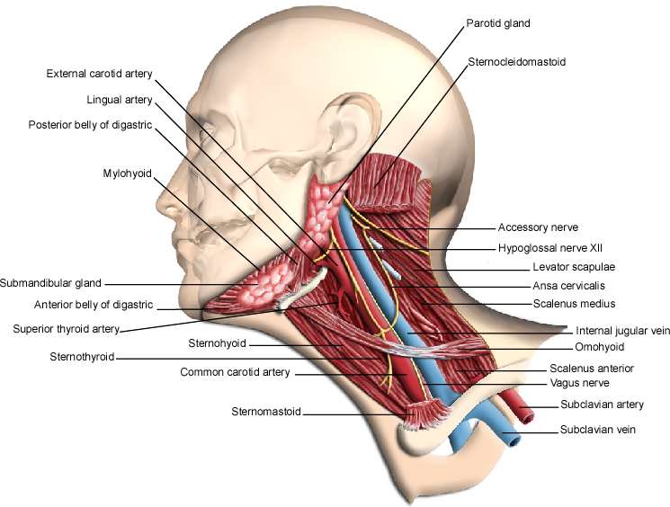
Lateral neck anatomy. This drawing shows muscles, glands, nerves and vessels of the neck. English labels.
This image by the Royal College of Surgeons of Ireland (RCSI) is retrieved from Health Education Assets Library (HEAL) of the University of Utah.
This image by the Royal College of Surgeons of Ireland (RCSI) is retrieved from Health Education Assets Library (HEAL) of the University of Utah.
Anatomical structures in item:
Uploaded by: rva
Netherlands, Leiden – Leiden University Medical Center, Leiden University
Collum
Arteria carotis externa
Arteria lingualis
Venter posterior musculus digastrici
Musculus digastricus
Musculus mylohyoideus
Glandula submandibularis
Venter posterior musculus digastrici anterior
Arteria thyroidea superior
Musculus sternothyroideus
Musculus sternohyoideus
Arteria carotis communis
Musculus sternocleidomastoideus
Vena subclavia
Arteria subclavia
Nervus vagus
Musculus scalenus anterior
Musculus omohyoideus
Vena jugularis interna
Musculus scalenus medius
Ansa cervicalis
Musculus levator scapulae
Nervus hypoglossus [XII]
Nervus accessorius [XI]
Glandula parotidea
Creator(s)/credit: Royal College of Surgeons of Ireland
Requirements for usage
You are free to use this item if you follow the requirements of the license:  View license
 View license
 If you use this item you should credit it as follows:
- For usage in print - copy and paste the line below:
- For digital usage (e.g. in PowerPoint, Impress, Word, Writer) - copy and paste the line below (optionally add the license icon):
"RCSI - Drawing Lateral neck anatomy - English labels" at AnatomyTOOL.org by Royal College of Surgeons of Ireland, license: Creative Commons Attribution-NonCommercial-ShareAlike


Comments