
nid: 62953
Additional formats:
None available
Description:
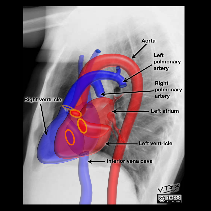
Position of heart and great vessels in chest x-ray: left lateral view. The anatomical position of several structures is projected on this chest x-ray. Other images, highlighting seperate structures can be found on Radiopaedia.org.
Case courtesy of Dr Vincent Tatco, Radiopaedia.org. From the case rID: 46331
Case courtesy of Dr Vincent Tatco, Radiopaedia.org. From the case rID: 46331
Anatomical structures in item:
Uploaded by: rva
Netherlands, Leiden – Leiden University Medical Center, Leiden University
Aorta
Arcus aortae
Arteria pulmonalis sinistra
Atrium dextrum
Vena cava inferior
Ventriculus sinister
Arteriae pulmonalis dextra
Truncus pulmonalis
Creator(s)/credit: Dr Vincent Tatco
Requirements for usage
You are free to use this item if you follow the requirements of the license:  View license
View license If you use this item you should credit it as follows:
- For usage in print - copy and paste the line below:
- For digital usage (e.g. in PowerPoint, Impress, Word, Writer) - copy and paste the line below (optionally add the license icon):
"Radiopaedia - Drawing/X-ray Position of heart and great vessels in chest x-ray: left lateral view - English labels" at AnatomyTOOL.org by Vincent Tatco, license: Creative Commons Attribution-NonCommercial-NoDerivs


Comments