nid: 60254
Additional formats:
None available
Description:
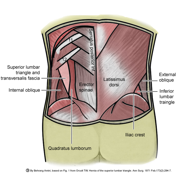
Superior and inferior lumbar triangle. This image shows the location of the superior and inferior lumbar triangle, through which lumbar hernias occur. English labels.
Case courtesy of Assoc Prof Frank Gaillard, Radiopaedia.org. From the case rID: 12934
Case courtesy of Assoc Prof Frank Gaillard, Radiopaedia.org. From the case rID: 12934
Anatomical structures in item:
Uploaded by: rva
Netherlands, Leiden – Leiden University Medical Center, Leiden University
Trigonum lumbale superius
Fascia transversalis
Musculus obliquus internus abdominis
Musculus erector spinae
Musculus serratus posterior inferior
Musculus quadratus lumborum
Crista iliaca
Trigonum lumbale inferius
Musculus obliquus externus abdominis
Creator(s)/credit: Dr Behrang Amini PhD, MD; Frank Gaillard MB.BS, MMed
Requirements for usage
You are free to use this item if you follow the requirements of the license:  View license
 View license
 If you use this item you should credit it as follows:
- For usage in print - copy and paste the line below:
- For digital usage (e.g. in PowerPoint, Impress, Word, Writer) - copy and paste the line below (optionally add the license icon):
"Radiopaedia - Drawing Superior and inferior lumbar triangle - English labels" at AnatomyTOOL.org by Behrang Amini and Frank Gaillard, license: Creative Commons Attribution-NonCommercial-ShareAlike. Based on Fig. 1 from Orcutt TW. Hernia of the superior lumbar triangle. Ann Surg. 1971 Feb;173(2):294-7
"Radiopaedia - Drawing Superior and inferior lumbar triangle - English labels" by Behrang Amini and Frank Gaillard, license: CC BY-NC-SA. Based on Fig. 1 from Orcutt TW. Hernia of the superior lumbar triangle. Ann Surg. 1971 Feb;173(2):294-7


Comments