nid: 60108
Additional formats:
None available
Description:
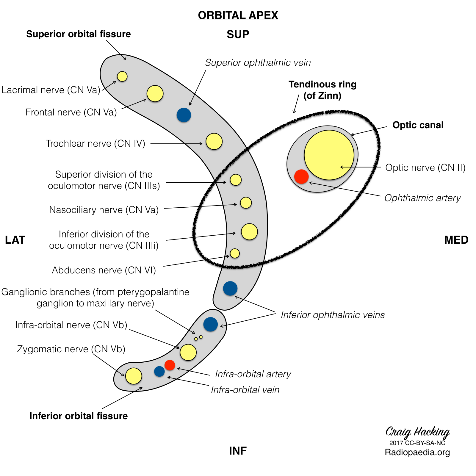
Nerves and vessels of orbital apex. The superior orbital fissure, inferior orbital fissure, optic canal, and tendinous ring are shown, through which the labeled nerves and vessels pass from the intracranial compartment into the orbit. English labels
Case courtesy of Assoc Prof Craig Hacking, Radiopaedia.org. From the case rID: 52363
Case courtesy of Assoc Prof Craig Hacking, Radiopaedia.org. From the case rID: 52363
Anatomical structures in item:
Uploaded by: rva
Netherlands, Leiden – Leiden University Medical Center, Leiden University
Fissura orbitalis superior
Zonula ciliaris
Canalis opticus
Nervus lacrimalis
Nervus frontalis
Nervus trochlearis [IV]
Ramus superior nervus oculomotorii
Nervus nasociliaris
Ramus inferior nervus oculomotorii
Nervus abducens [VI]
Rami ganglionares nervus maxillaris ad ganglion pterygopalatinum
Nervus infraorbitalis
Nervus zygomaticus
Fissura orbitalis inferior
Vena ophthalmica inferior
Arteria ophthalmica
Nervus opticus
Vena ophthalmica superior
Creator(s)/credit: Craig Hacking MB.BS, BSc
Requirements for usage
You are free to use this item if you follow the requirements of the license:  View license
 View license
 If you use this item you should credit it as follows:
- For usage in print - copy and paste the line below:
- For digital usage (e.g. in PowerPoint, Impress, Word, Writer) - copy and paste the line below (optionally add the license icon):
"Radiopaedia - Drawing Nerves and vessels of orbital apex - English labels" at AnatomyTOOL.org by Craig Hacking, license: Creative Commons Attribution-NonCommercial-ShareAlike


Comments