
nid: 60242
Additional formats:
None available
Description:
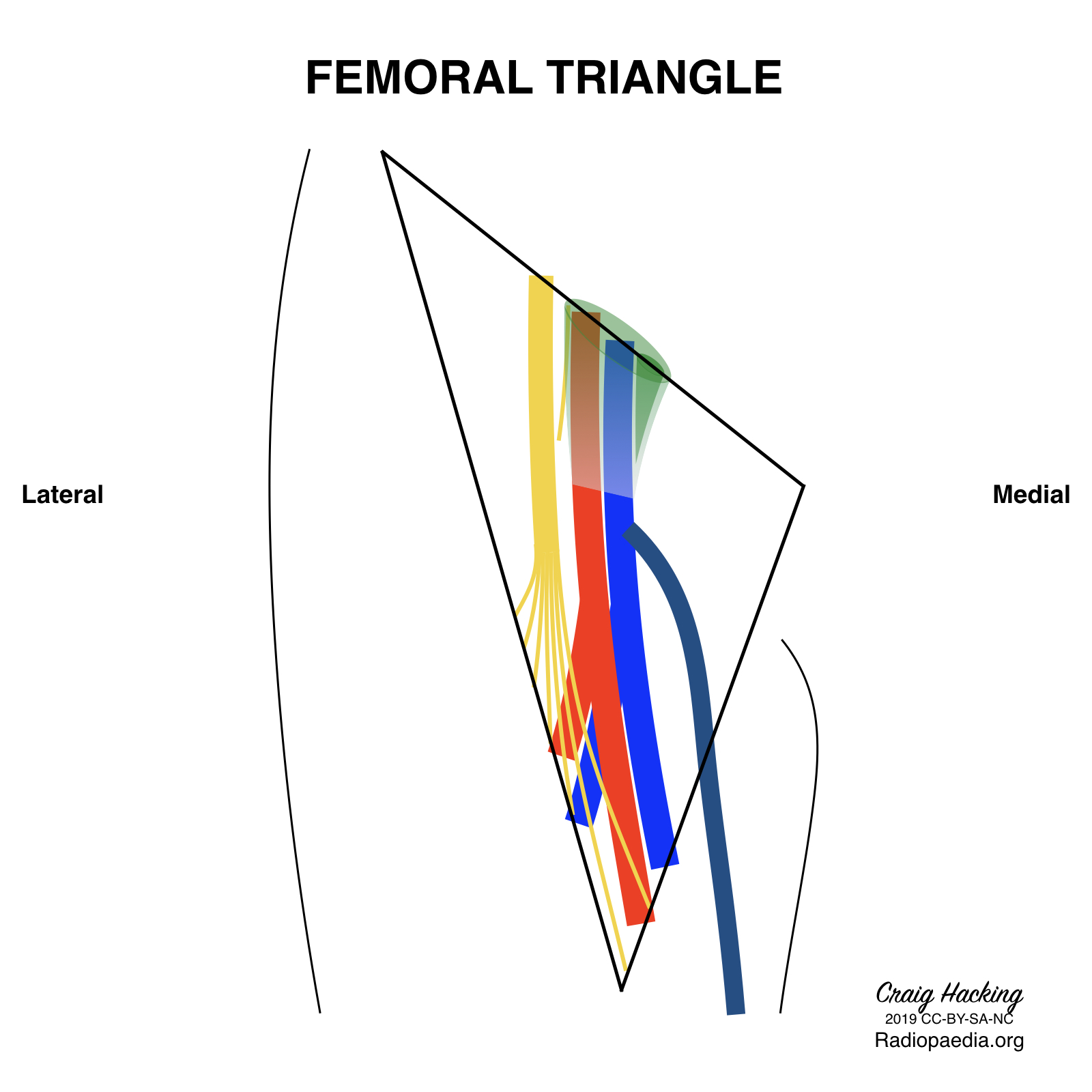
Femoral triangle schematic. The femoral triangle is an anatomical space in the upper thigh, it's borders are the sartorius muscle (lateral), adductor longus muscle (medial), inguinal ligament (superior), and illopsoas and pectineus muscle (floor). The contents are the femoral nerve, artery, vein and canal. No labels
Case courtesy of Assoc Prof Craig Hacking, Radiopaedia.org. From the case rID: 70536
Case courtesy of Assoc Prof Craig Hacking, Radiopaedia.org. From the case rID: 70536
Anatomical structures in item:
Uploaded by: rva
Netherlands, Leiden – Leiden University Medical Center, Leiden University
Femoral triangle
Musculus sartorius
Musculus adductor longus
Ramus femoralis nervus genitofemoralis
Nervus femoralis
Arteria femoralis
superficial femoral artery
Arteria profunda femoris
Vena profunda femoris
Vena saphena magna
Vena femoralis
Vena femoralis
Canalis femoralis
Anulus femoralis
Femoral sheath
Ligamentum inguinale
Creator(s)/credit: Craig Hacking MB.BS, BSc
Requirements for usage
You are free to use this item if you follow the requirements of the license:  View license
View license If you use this item you should credit it as follows:
- For usage in print - copy and paste the line below:
- For digital usage (e.g. in PowerPoint, Impress, Word, Writer) - copy and paste the line below (optionally add the license icon):
"Radiopaedia - Drawing Femoral triangle schematic - no labels" at AnatomyTOOL.org by Craig Hacking, license: Creative Commons Attribution-NonCommercial-ShareAlike


Comments