
nid: 60116
Additional formats:
None available
Description:
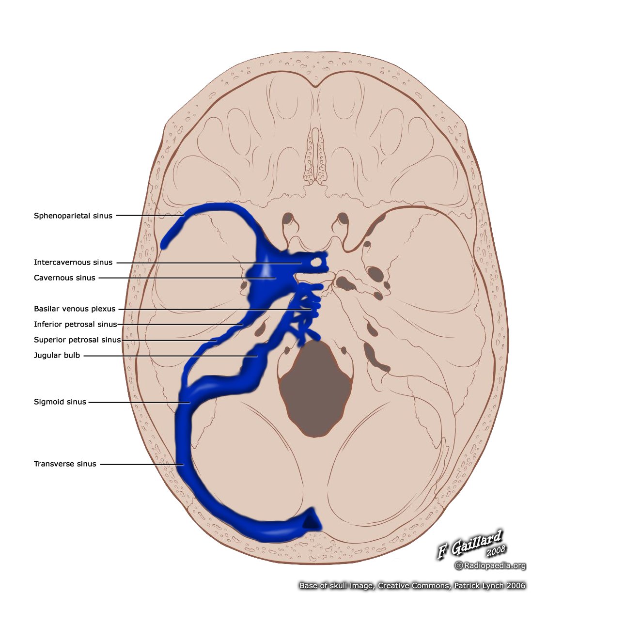
Dural venous sinuses. The dural venous sinuses are located between the endosteal layer and meningeal layer of the dura mater and receive blood from the cerebral veins and cerebrospinal fluid via arachnoid granulations. English labels
Case courtesy of Assoc Prof Frank Gaillard, Radiopaedia.org. From the case rID: 36180
Case courtesy of Assoc Prof Frank Gaillard, Radiopaedia.org. From the case rID: 36180
Anatomical structures in item:
Uploaded by: rva
Netherlands, Leiden – Leiden University Medical Center, Leiden University
Sinus sphenoparietalis
Sinus intercavernosus anterior
Sinus cavernosus
Plexus venosus basilaris
Basis cranii interna
Basis cranii
Sinus petrosus inferior
Sinus petrosus superior
Sinus sigmoideus
Sinus transversus
Creator(s)/credit: Frank Gaillard MB.BS, MMed; Patrick J. Lynch, medical illustrator
Requirements for usage
You are free to use this item if you follow the requirements of the license:  View license
View license If you use this item you should credit it as follows:
- For usage in print - copy and paste the line below:
- For digital usage (e.g. in PowerPoint, Impress, Word, Writer) - copy and paste the line below (optionally add the license icon):
"Radiopaedia - Drawing Dural venous sinuses - English labels" at AnatomyTOOL.org by Frank Gaillard and Patrick J. Lynch, license: Creative Commons Attribution-NonCommercial-ShareAlike
"Radiopaedia - Drawing Dural venous sinuses - English labels" by Frank Gaillard and Patrick J. Lynch, license: CC BY-NC-SA


Comments