
nid: 62458
Additional formats:
None available
Description:
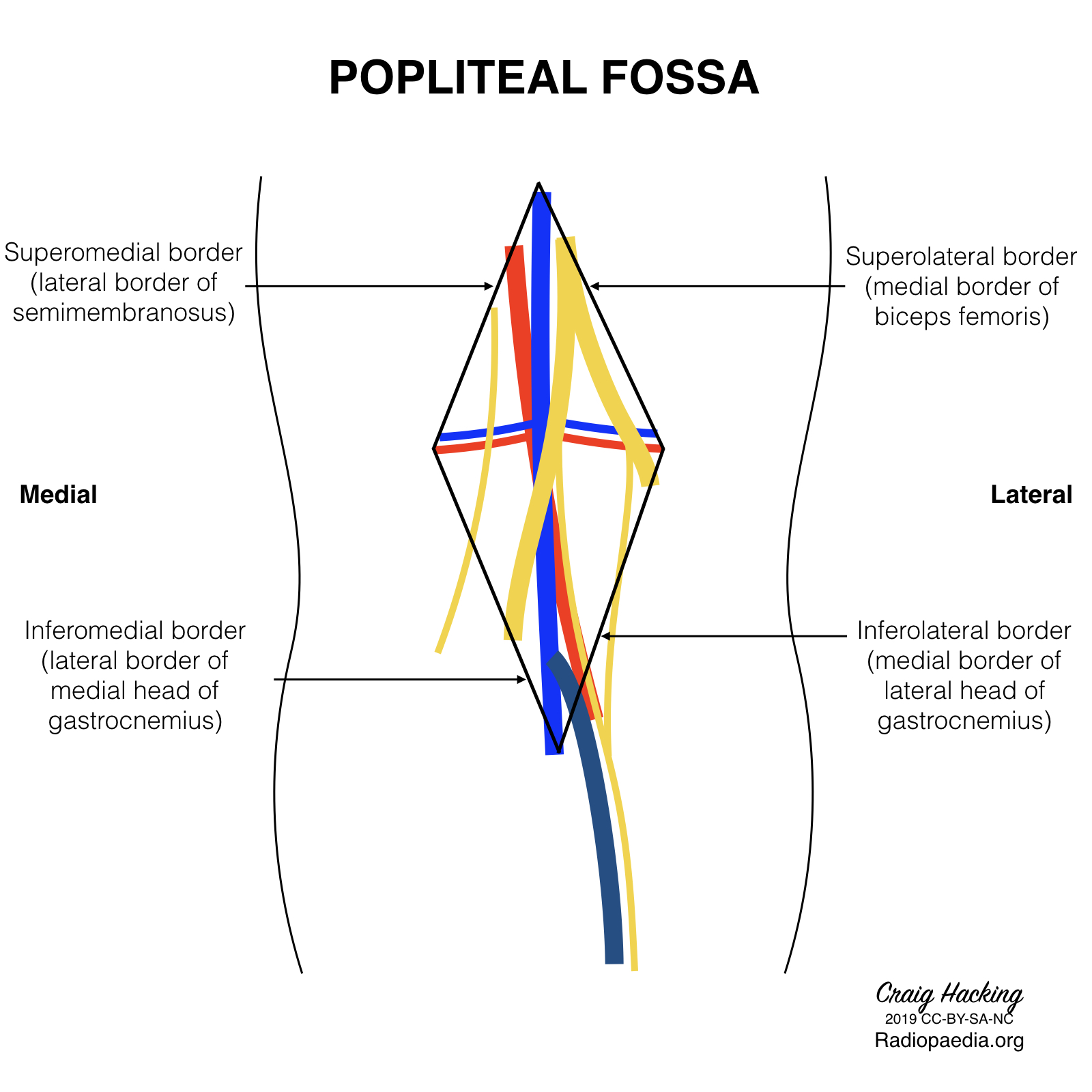
Boundaries of the popliteal fossa. The boundaries of the popliteal fossa are created by the semimembranosa muscle, the medial and lateral head of the gastrocnemius muscle, and the biceps femoris muscle. English labels.
Case courtesy of Assoc Prof Craig Hacking, Radiopaedia.org. From the case rID: 70604
Case courtesy of Assoc Prof Craig Hacking, Radiopaedia.org. From the case rID: 70604
Anatomical structures in item:
Uploaded by: rva
Netherlands, Leiden – Leiden University Medical Center, Leiden University
Fossa poplitea
Creator(s)/credit: Craig Hacking MB.BS, BSc
Requirements for usage
You are free to use this item if you follow the requirements of the license:  View license
View license If you use this item you should credit it as follows:
- For usage in print - copy and paste the line below:
- For digital usage (e.g. in PowerPoint, Impress, Word, Writer) - copy and paste the line below (optionally add the license icon):
"Radiopaedia - Drawing Boundaries of the popliteal fossa - English labels" at AnatomyTOOL.org by Craig Hacking, license: Creative Commons Attribution-NonCommercial-ShareAlike


Comments