
nid: 58137
Additional formats:
None available
Description:
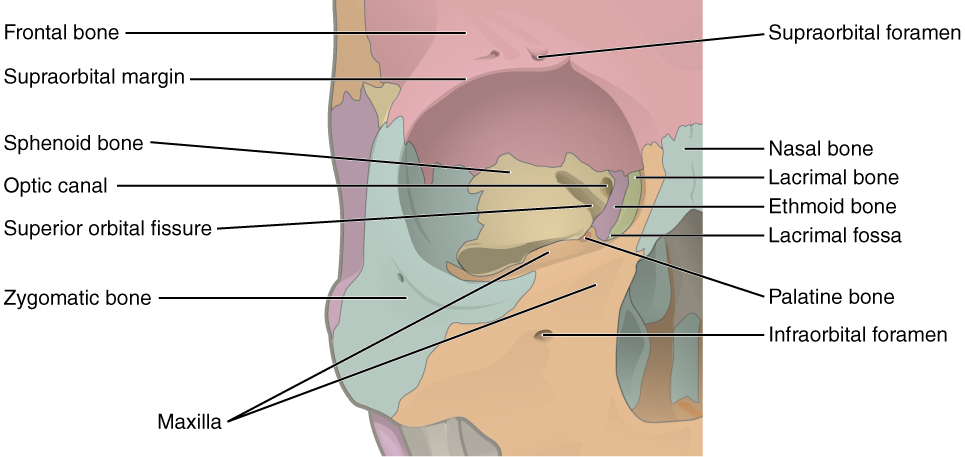
Seven skull bones contribute to the walls of the orbit. Opening into the posterior orbit from the cranial cavity are the optic canal and superior orbital fissure. English labels. From OpenStax book 'Anatomy and Physiology', fig. 7.19.
Anatomical structures in item:
Uploaded by: Jorn IJkhout
Netherlands, Leiden – Leiden University Medical Center, Leiden University
Bone
Cranium
Orbita
Maxilla
Os zygomaticum
Os frontale
Os ethmoidale
Os lacrimale
Os sphenoidale
Os nasale
Os palatinum
Fissura orbitalis superior
Canalis opticus
Creator(s)/credit: OpenStax
Requirements for usage
You are free to use this item if you follow the requirements of the license:  View license
View license If you use this item you should credit it as follows:
- For usage in print - copy and paste the line below:
- For digital usage (e.g. in PowerPoint, Impress, Word, Writer) - copy and paste the line below (optionally add the license icon):
"OpenStax AnatPhys fig.7.19 - Bones Forming Orbit - English labels" at AnatomyTOOL.org by OpenStax, license: Creative Commons Attribution. Source: book 'Anatomy and Physiology', https://openstax.org/details/books/anatomy-and-physiology.
"OpenStax AnatPhys fig.7.19 - Bones Forming Orbit - English labels" by OpenStax, license: CC BY. Source: book 'Anatomy and Physiology', https://openstax.org/details/books/anatomy-and-physiology.


Comments